You have no items in your shopping cart.
Cart summary
Item 1 of 5
Item 1 of 5
VIM Antibody
Catalog Number: orb1264636
| Catalog Number | orb1264636 |
|---|---|
| Category | Antibodies |
| Description | VIM Antibody |
| Species/Host | Rabbit |
| Clonality | Polyclonal |
| Tested applications | FC, IF, IHC-P, WB |
| Predicted Reactivity | Bovine, Gallus, Hamster, Monkey, Mouse, Porcine, Rat, Xenopus |
| Reactivity | Human |
| Isotype | Rabbit Ig |
| Immunogen | This Vimentin antibody is generated from rabbits immunized with a KLH conjugated synthetic peptide between 430-457 amino acids from the C-terminal region of human Vimentin. |
| Concentration | batch dependent |
| Dilution range | For WB starting dilution is: 1:1000For IF starting dilution is: 1:100For IHC-P starting dilution is: 1:10~50For FACS starting dilution is: 1:10~50 |
| Form/Appearance | Liquid |
| Conjugation | Unconjugated |
| MW | 54 kDa |
| Target | VIM |
| UniProt ID | P08670 |
| NCBI | P08670 |
| Storage | Store at 4°C for three months and -20°C, stable for up to one year. As with all antibodies care should be taken to avoid repeated freeze thaw cycles. Antibodies should not be exposed to prolonged high temperatures. |
| Buffer/Preservatives | Supplied in PBS with 0.09% (W/V) sodium azide. |
| Alternative names | Vimentin, VIM Read more... |
| Note | For research use only |
| Application notes | For WB starting dilution is: 1:1000For IF starting dilution is: 1:100For IHC-P starting dilution is: 1:10~50For FACS starting dilution is: 1:10~50 |
| Expiration Date | 12 months from date of receipt. |
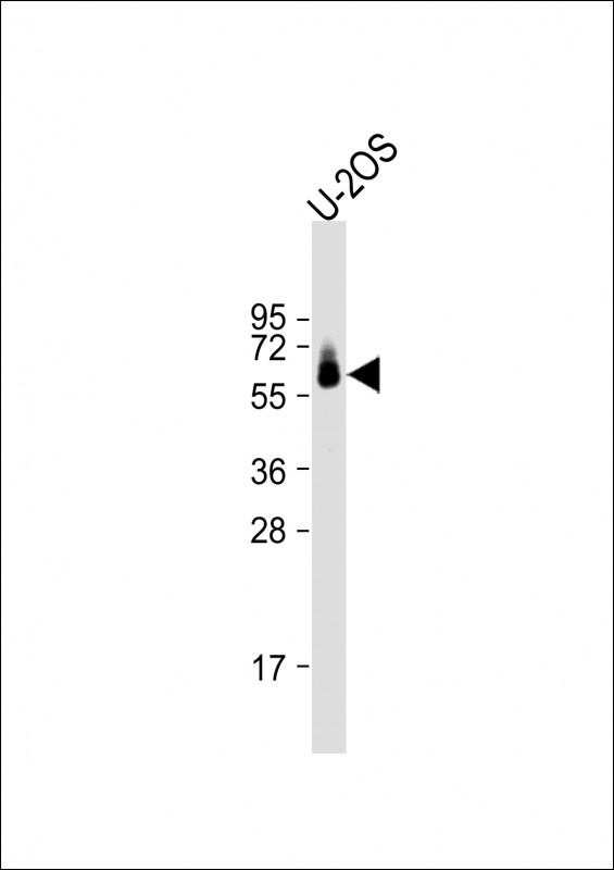
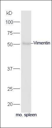
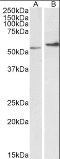
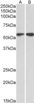
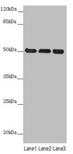
Western Blot at 1:1000 dilution + U-2OS whole cell lysate Lysates/proteins at 20 ug per lane.
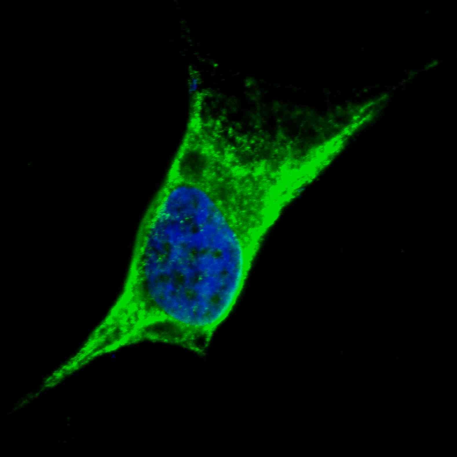
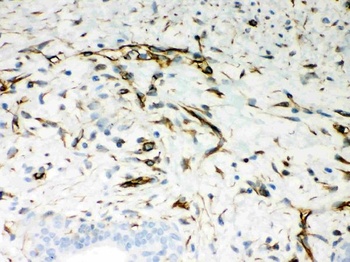
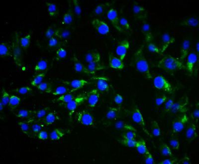
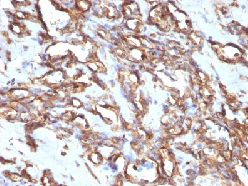
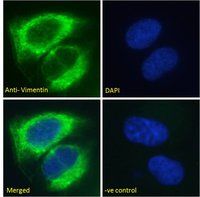
Fluorescent confocal image of SY5Y cells stained with Vimentin antibody. SY5Y cells were fixed with 4% PFA (20 min), permeabilized with Triton X-100 (0.2%, 30 min). Cells were then incubated with Antibody Vimentin primary antibody (1:100, 2 h at room temperature). For secondary antibody, Alexa Fluor 488 conjugated donkey anti-rabbit antibody (green) was used (1:1000, 1h). Nuclei were counterstained with Hoechst 33342 (blue) (10 ug/ml, 5 min). Note the highly specific localization of the Vimentin immunosignal to the cytoskeleton, supported by Human Protein Atlas Data (http://www.proteinatlas.org/ENSG00000026025).
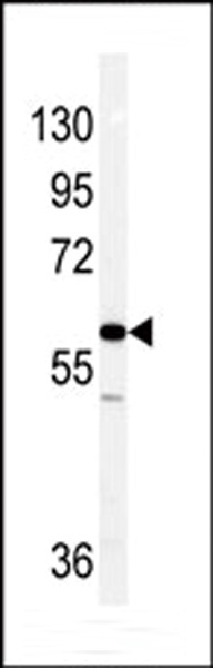
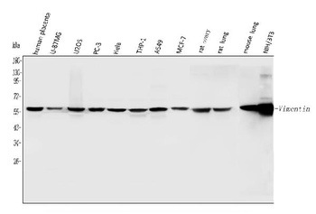
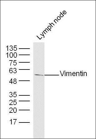
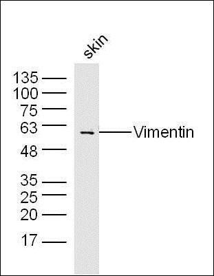
Western blot analysis of Vimentin antibody in NCI-H460 cell line lysates (35 ug/lane).
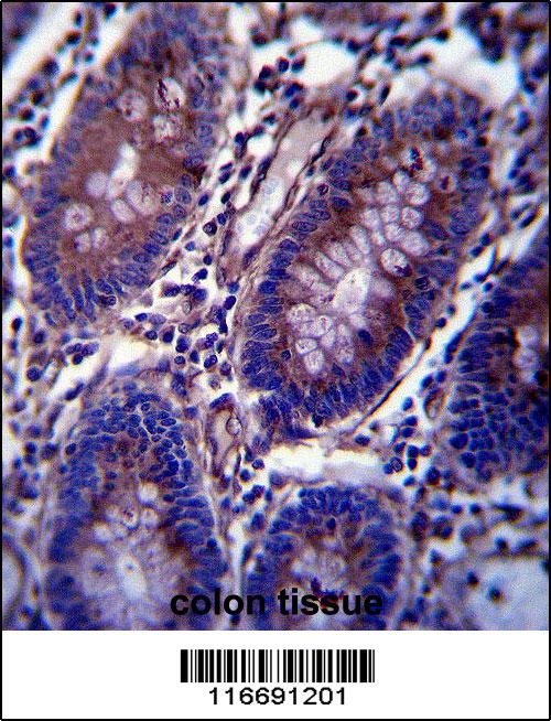
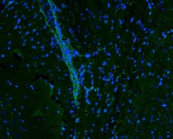
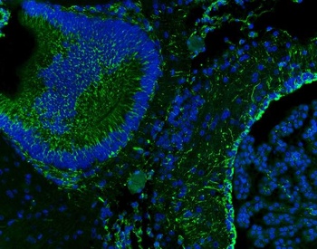
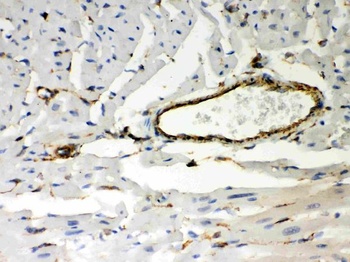
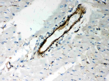
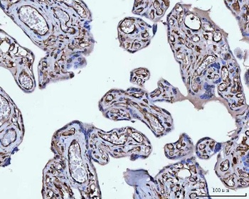
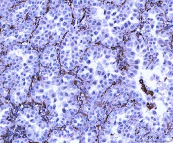
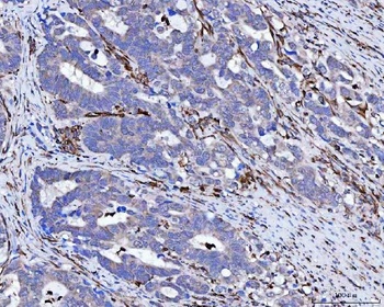
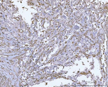
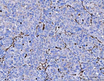
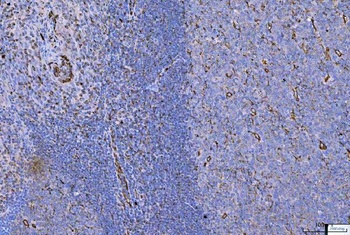
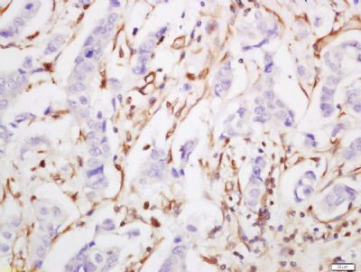
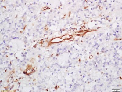
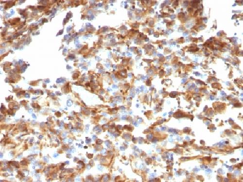
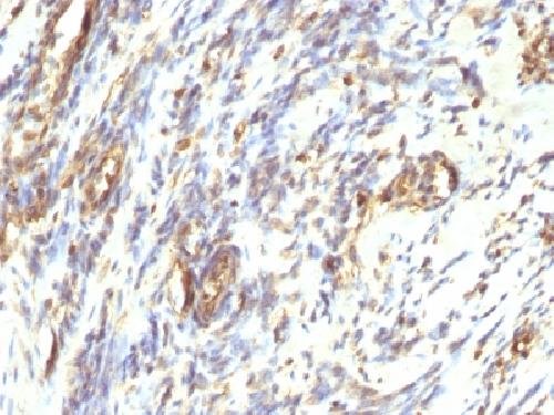
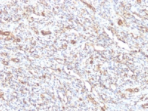
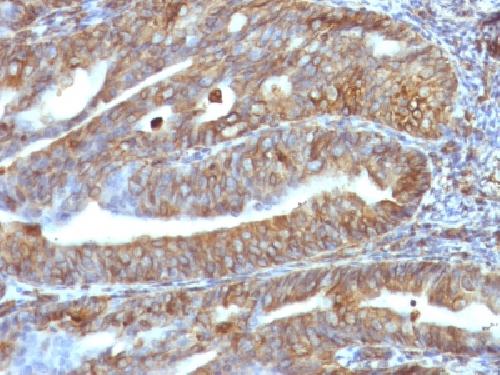
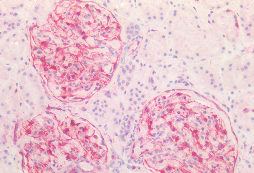
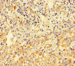
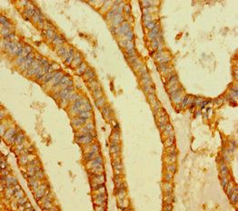
Vimentin Antibody immunohistochemistry analysis in formalin fixed and paraffin embedded human colon tissue followed by peroxidase conjugation of the secondary antibody and DAB staining.
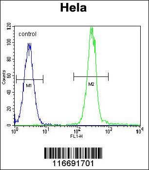
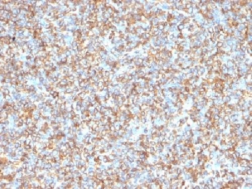
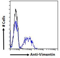
Flow cytometric analysis of Hela cells (right histogram) compared to a negative control cell (left histogram). FITC-conjugated goat-anti-rabbit secondary antibodies were used for the analysis.
- Item 1 of 12
Vimentin Antibody [orb251542]
IF, IHC, WB
Hamster
Human, Mouse, Rat
Rabbit
Polyclonal
Unconjugated
10 μg, 100 μg - Item 1 of 7
Vimentin antibody [orb158714]
FC, ICC, IF, WB
Bovine, Canine, Equine, Porcine, Rat
Human, Mouse, Rabbit
Rabbit
Polyclonal
Unconjugated
200 μl, 50 μl, 100 μl - Item 1 of 6
- Item 1 of 5
Vimentin antibody [orb233645]
ELISA, FC, IF, IHC, WB
Bovine, Canine, Human, Mouse, Porcine, Rat
Goat
Polyclonal
Unconjugated
100 μg - Item 1 of 4
Submit a review
Filter by Rating

- 5 stars

- 4 stars
- 3 stars
- 2 stars
- 1 stars