You have no items in your shopping cart.
Cart summary
Item 1 of 10
Item 1 of 10
VCP Antibody
Catalog Number: orb259631
| Catalog Number | orb259631 |
|---|---|
| Category | Antibodies |
| Description | VCP Antibody |
| Species/Host | Rabbit |
| Clonality | Polyclonal |
| Tested applications | FC, ICC, IF, IHC, IHC-Fr, WB |
| Predicted Reactivity | Hamster |
| Reactivity | Human, Mouse, Rat |
| Isotype | Rabbit IgG |
| Immunogen | A synthetic peptide corresponding to a sequence at the C-terminus of human VCP (732-760aa RRDHFEEAMRFARRSVSDNDIRKYEMFAQ), identical to the related rat and mouse sequences. |
| Concentration | Adding 0.2 ml of distilled water will yield a concentration of 500 μg/ml. |
| Dilution range | Western blot, 0.1-0.5μg/ml, Human, Mouse, Rat Immunohistochemistry (Paraffin-embedded Section), 0.5-1μg/ml, Human, Mouse, Rat, By Heat Immunohistochemistry (Frozen Section), 0.5-1μg/ml, Mouse, Rat, Immunocytochemistry/Immunofluorescence, 2μg/ml, Human Flow Cytometry, 1-3μg/1x106 cells, Human |
| Form/Appearance | Lyophilized |
| Conjugation | Unconjugated |
| MW | 89322 MW |
| UniProt ID | P55072 |
| Storage | Store at -20˚C for one year from date of receipt. After reconstitution, at 4˚C for one month. It can also be aliquotted and stored frozen at -20˚C for six months. Avoid repeated freeze-thaw cycles. |
| Alternative names | Transitional endoplasmic reticulum ATPase;TER ATPa Read more... |
| Note | For research use only |
| Application notes | WB: The detection limit for VCP is approximately 0.1ng/lane under reducing conditions. Tested Species: In-house tested species with positive results. By Heat: Boiling the paraffin sections in 10mM citrate buffer, pH6.0, for 20mins is required for the staining of formalin/paraffin sections. Other applications have not been tested. Optimal dilutions should be determined by end users. . Add 0.2ml of distilled water will yield a concentration of 500ug/ml. |
| Expiration Date | 12 months from date of receipt. |
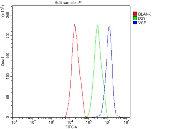
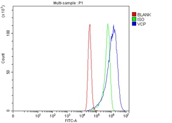
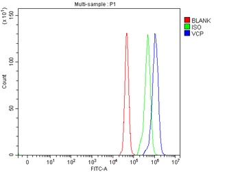
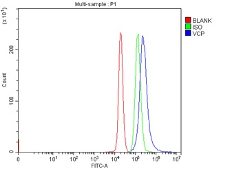
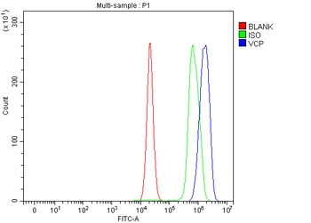
Flow Cytometry analysis of HepG2 cells using anti-VCP antibody (Blue line).Isotype control antibody (Green line) was rabbit IgG .Unlabelled sample (Red line) was also used as a control.
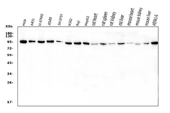
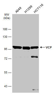
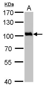
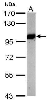
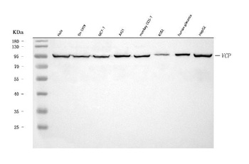
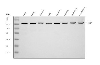
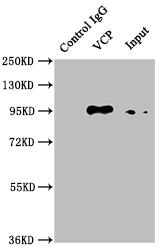
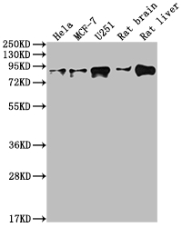
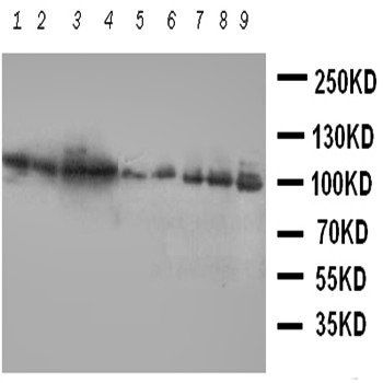
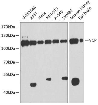
WB:1:HeLa;2:A431;3:U-87MG;4:A549;5:SH-SY5Y cell;6:K562 cell;7:Raji cell;8:HepG2 cell;9:rat heart tissue;10:rat spleen tissue;11:rat kidney tissue;12:rat liver tissue;13:mouse heart tissue;14:mouse kidney tissue;15:mouse liver tissue;16:HEPA1-6 cell.
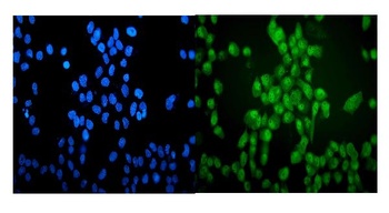
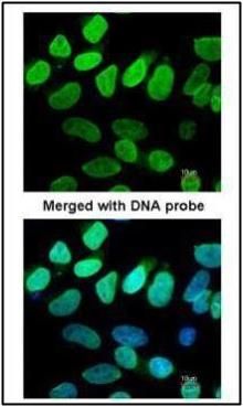
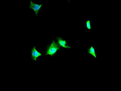
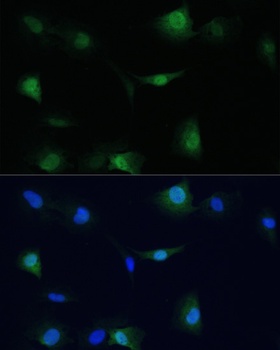
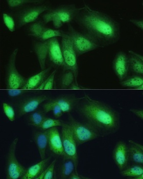
IF analysis of VCP using anti-VCP antibody. VCP was detected in immunocytochemical section of A431 cells.
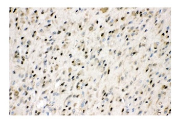
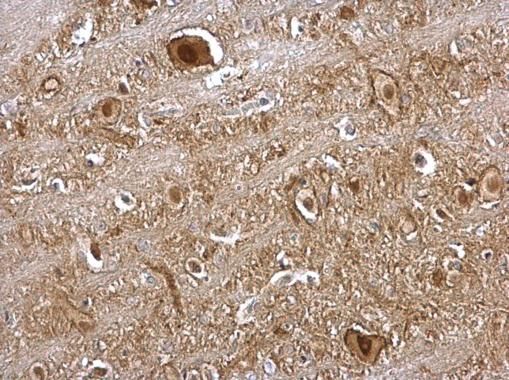
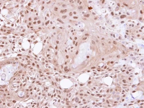
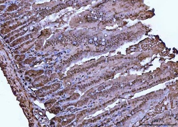
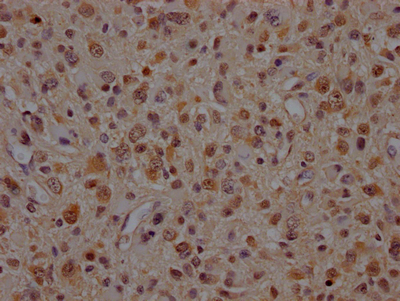
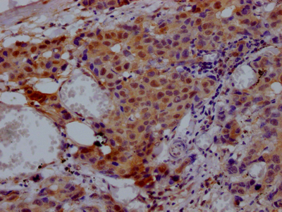
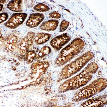
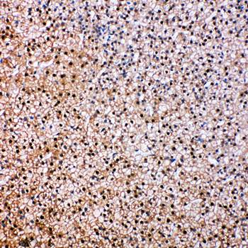
IHC analysis of VCP using anti-VCP antibody. VCP was detected in paraffin-embedded section of human glioma tissue.
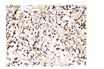
IHC analysis of VCP using anti-VCP antibody. VCP was detected in paraffin-embedded section of human glioma tissue.
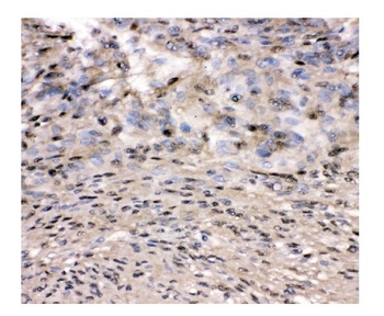
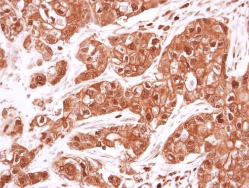
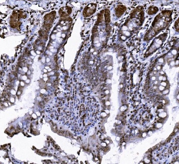
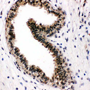
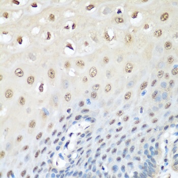
IHC analysis of VCP using anti-VCP antibody. VCP was detected in paraffin-embedded section of human meningeoma tissue.
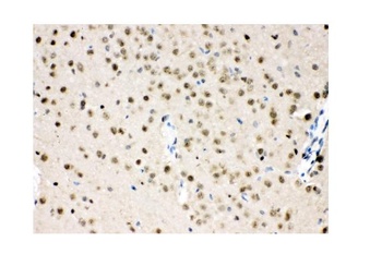
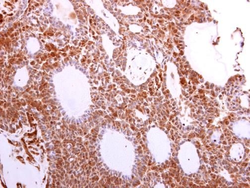
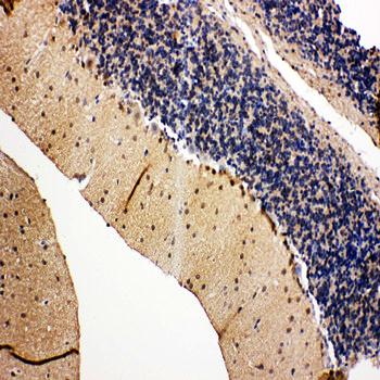
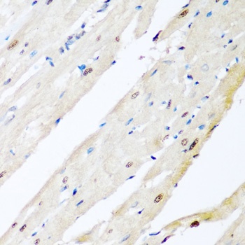
IHC analysis of VCP using anti-VCP antibody. VCP was detected in paraffin-embedded section of mouse brain tissue.
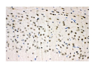
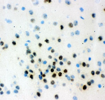
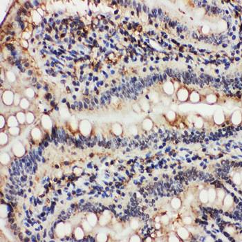
IHC analysis of VCP using anti-VCP antibody. VCP was detected in paraffin-embedded section of rat brain tissue.
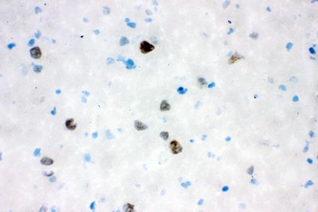
IHC analysis of VCP using anti-VCP antibody. VCP was detected in frozen section of mouse brain tissue.
IHC analysis of VCP using anti-VCP antibody. VCP was detected in frozen section of rat brain tissue.
- Item 1 of 8
VCP antibody [orb556833]
ICC, IHC, IHC-P, IP, WB
Canine, Human, Mouse, Rat, Zebrafish
Rabbit
Polyclonal
Unconjugated
100 μl - Item 1 of 8
VCP Antibody [orb1289976]
ELISA, FC, IHC, WB
Human, Monkey, Mouse, Rat
Rabbit
Polyclonal
Unconjugated
100 μg, 10 μg - Item 1 of 5
- Item 1 of 6
VCP Antibody [orb107641]
FC, ICC, IF, IHC, IHC-Fr, WB
Hamster
Human, Monkey, Mouse, Rat
Rabbit
Polyclonal
Unconjugated
10 μg, 100 μg - Item 1 of 5
Submit a review
Filter by Rating

- 5 stars

- 4 stars
- 3 stars
- 2 stars
- 1 stars