You have no items in your shopping cart.
Description
Research Area
Cell Biology
Images & Validation
−Item 1 of 6
| Tested Applications | FC, IF, IHC-P, WB |
|---|---|
| Dilution Range | IF - 1:10-50, WB - 1:1000, IHC-P - 1:25, FC - 1:10-50 |
| Reactivity | Human, Mouse, Rat |
| Predicted Reactivity | Equine, Porcine |
Key Properties
−| Host | Rabbit |
|---|---|
| Clonality | Polyclonal |
| Isotype | Rabbit IgG |
| Immunogen | This UCHL1 antibody is generated from rabbits immunized with a KLH conjugated synthetic peptide between 187-216 amino acids from the C-terminal region of human UCHL1. Antigen Region: 187-216 aa. |
| Target | UCHL1 |
| Molecular Weight | 24824 Da |
| Conjugation | Unconjugated |
Storage & Handling
−| Storage | Maintain refrigerated at 2-8°C for up to 2 weeks. For long term storage store at -20°C in small aliquots to prevent freeze-thaw cycles |
|---|---|
| Form/Appearance | Purified polyclonal antibody supplied in PBS with 0.09% (W/V) sodium azide. This antibody is purified through a protein A column, followed by peptide affinity purification. |
| Expiration Date | 12 months from date of receipt. |
| Disclaimer | For research use only |
Alternative Names
−Ubiquitin carboxyl-terminal hydrolase isozyme L1, UCH-L1, 6---, Neuron cytoplasmic protein 95, PGP 95, PGP95, Ubiquitin thioesterase L1, UCHL1
Similar Products
−- Item 1 of 4
UCHL1 Antibody (C-term) [orb1931712]
IF, IHC-P, WB
Equine, Mouse, Porcine
Human, Rat
Rabbit
Polyclonal
Unconjugated
50 μl, 100 μl - Item 1 of 3
UCHL1 Antibody (C-term) [orb1927319]
IHC-P, WB
Equine, Mouse, Porcine
Human, Rat
Mouse
Monoclonal
Unconjugated
50 μl, 100 μl - Item 1 of 3
UCHL1 Antibody (C-term) [orb1788350]
WB
Human, Mouse, Rat
Rabbit
Polyclonal
Unconjugated
- Item 1 of 2
UCHL1 Antibody (C-term) [orb1788247]
WB
Equine, Porcine
Human, Mouse, Rat
Rabbit
Polyclonal
Unconjugated
- Item 1 of 1
UCHL1 Antibody (C-term) [orb1788261]
WB
Human, Mouse, Rat
Mouse
Monoclonal
Unconjugated
Quality Guarantee
Explore bioreagents carefree to elevate your research. All our products are rigorously tested for performance. If a product does not perform as described on its datasheet, our scientific support team will provide expert troubleshooting, a prompt replacement, or a refund. For full details, please see our Terms & Conditions and Buying Guide. Contact us at [email protected].
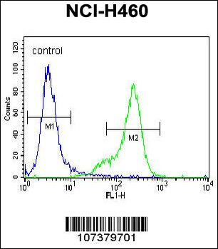
UCHL1 Antibody (C-term) flow cytometric analysis of NCI-H460 cells (right histogram) compared to a negative control cell (left histogram). FITC-conjugated goat-anti-rabbit secondary antibodies were used for the analysis.
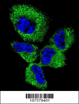
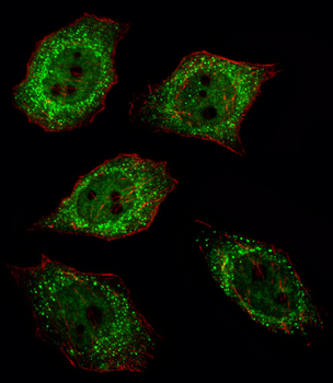
Confocal immunofluorescent analysis of UCHL1 Antibody (C-term) with NCI-H460 cell followed by Alexa Fluor 488-conjugated goat anti-rabbit lgG (green). DAPI was used to stain the cell nuclear (blue).
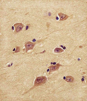
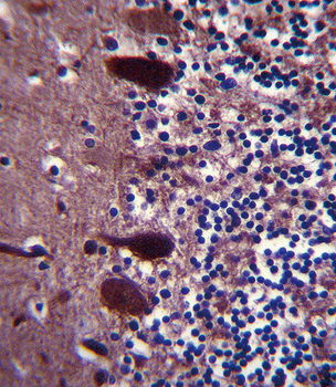
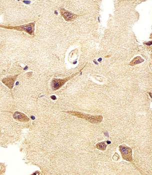
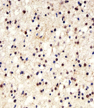
Staining UCHL1 in human brain tissue sections by Immunohistochemistry (IHC-P - paraformaldehyde-fixed, paraffin-embedded sections). Tissue was fixed with formaldehyde and blocked with 3% BSA for 0.5 hour at room temperature; antigen retrieval was by heat mediation with a citrate buffer (pH6). Samples were incubated with primary antibody (1/25) for 1 hours at 37°C. A undiluted biotinylated goat polyvalent antibody was used as the secondary antibody.

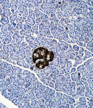
Staining UCHL1 in human lung adenocarcinoma tissue sections by Immunohistochemistry (IHC-P - paraformaldehyde-fixed, paraffin-embedded sections). Tissue was fixed with formaldehyde and blocked with 3% BSA for 0.5 hour at room temperature; antigen retrieval was by heat mediation with a citrate buffer (pH6). Samples were incubated with primary antibody (1/25) for 1 hours at 37°C. A undiluted biotinylated goat polyvalent antibody was used as the secondary antibody.
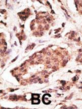
Formalin-fixed and paraffin-embedded human cancer tissue reacted with the primary antibody, which was peroxidase-conjugated to the secondary antibody, followed by DAB staining. This data demonstrates the use of this antibody for immunohistochemistry; clinical relevance has not been evaluated. BC = breast carcinoma; HC = hepatocarcinoma.
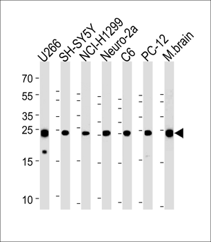
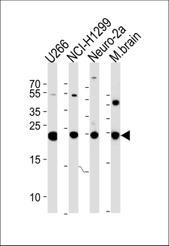
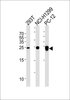
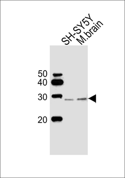
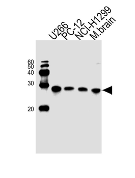
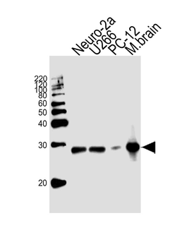
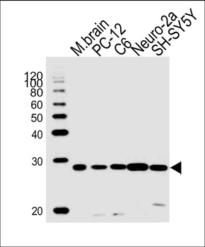
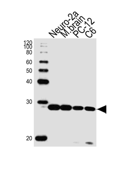
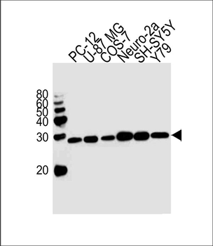
UCHL1 Antibody (C-term) western blot analysis in U266, SH-SY5Y, NCI-H1299, mouse Neuro-2a, rat C6, PC-12 cell line and mouse brain tissue lysates (35 ug/lane). This demonstrates the UCHL1 antibody detected the UCHL1 protein (arrow).
Quick Database Links
UniProt Details
− No UniProt data available
NCBI Reference Sequences
−Associated Accession Numbers
Curated reference sequences for the gene transcript and protein product| Protein | NP_004172.2 |
|---|
Documents Download
Datasheet
Product Information
Request a Document
Protocol Information
WB
Western Blot (IB, immunoblot)
IHC-P
Immunohistochemistry Paraffin
FC
Flow Cytometry
IF
Immunofluorescence
UCHL1 Antibody (C-term) (orb1931713)

- 0.0
Based on 0 reviews
Participating in our Biorbyt product reviews program enables you to support fellow scientists by sharing your firsthand experience with our products.
Login to Submit a ReviewAvailable Sizes
Select a size below
Choose Conjugation or Carrier Free Version
Free Secondary Antibody (20 ul)0/0
Please add an antibody product to your cart first.