You have no items in your shopping cart.
Cart summary
Item 1 of 2
Item 1 of 2
Ubiquitin Activating Enzyme E1 Antibody
Catalog Number: orb344629

| Catalog Number | orb344629 |
|---|---|
| Category | Antibodies |
| Description | Ubiquitin Activating Enzyme E1 antibody |
| Clonality | Polyclonal |
| Species/Host | Rabbit |
| Isotype | IgG |
| Conjugation | Unconjugated |
| Reactivity | Human |
| Form/Appearance | Lyophilized |
| Concentration | 1.2 mg/mL |
| Buffer/Preservatives | 0.01% (w/v) Sodium Azide |
| Purity | This antibody is directed against human Ubiquitin Activating Enzyme E1 protein. The product was protein A purified from monospecific antiserum followed by further purification to remove the GST tag. A BLAST analysis was used to suggest that this antibody would react with Ubiquitin Activating Enzyme E1 protein from human (100%) rabbit (96%), mouse (95%), rat (95%) and dog (93%) based on a high degree of sequence homology. Cross reactivity against this protein from other sources has not been determined. |
| Immunogen | Anti-Ubiquitin Activating Enzyme E1 antibody was prepared from whole rabbit serum produced by repeated immunizations with a recombinant protein corresponding to full length Human Ubiquitin Activating Enzyme E1. |
| UniProt ID | P22314 |
| Tested applications | ELISA, IHC, WB |
| Dilution range | ELISA: 1:2,000 - 1:10,000, IHC: 2 mg/ml - 20 µg/ml, WB: 1:1,000 - 1:5,000 |
| Application notes | This purified antibody has been tested for use in ELISA, immunohistochemistry and western blot. Specific conditions for reactivity should be optimized by the end user. Expect a band at ~118 kDa in size corresponding to UBE1 by western blotting in the appropriate cell lysate or extract. |
| Antibody Type | Primary Antibody |
| Storage | Store vial at 4° C prior to restoration. For extended storage aliquot contents and freeze at -20° C or below. Avoid cycles of freezing and thawing. Centrifuge product if not completely clear after standing at room temperature. This product is stable for several weeks at 4° C as an undiluted liquid. Dilute only prior to immediate use. |
| Alternative names | rabbit anti-Ubiquitin Activating Enzyme E1 Antibod Read more... |
| Note | For research use only |
| NCBI | 23510338 |
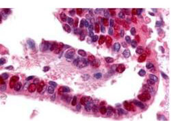
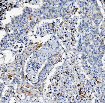
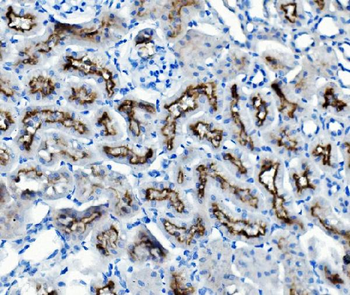
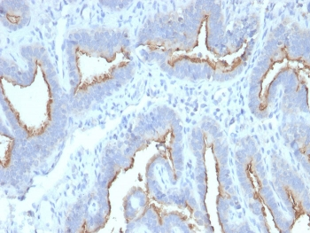
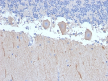
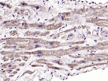
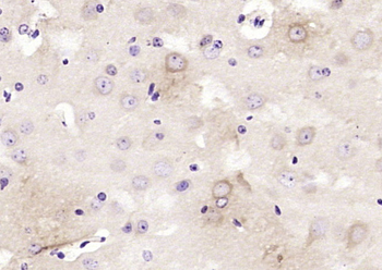
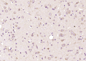
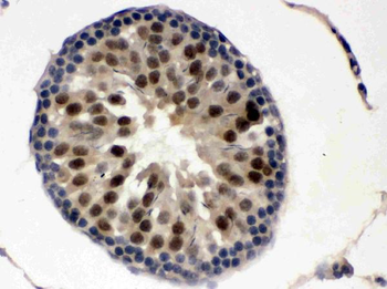
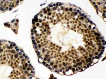
Biorbyt's Affinity Purified anti-Ubiquitin Activating Enzyme antibody was used at a 10 µg/ml to detect UBE1 in a variety of tissues including adrenal, breast, colon (epithelium), kidney, liver, lung (respiratory epithelium), ovary (oocyte and endothelium), pancreas (islet and exocrine), placenta, prostate (epithelium), skin (epithelium), spleen (lymphocytes), stomach (chief), testis, thymus, tonsil, and uterus (glandular, stroma). In many cells a punctate nuclear staining was observed. Other cells showed both cytoplasmic and nuclear staining. This image shows UBE1 staining of human lung tissue. Tissue was formalin-fixed and paraffin embedded.
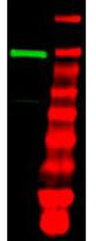
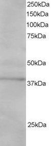
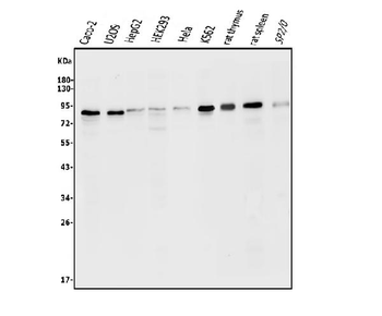
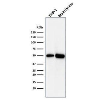
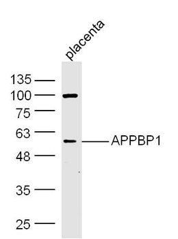
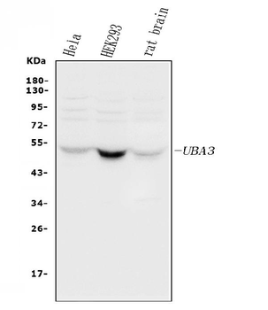
Western blot using Biorbyt's purified anti-Ubiquitin Activating Enzyme (E1) antibody shows detection of a band at ~118 kDa corresponding to UBE1 (lane 1 800 nm channel). Approximately 35 µg of an A431 whole cell lysate (p/n orb348665) was separated on a 4-20% Tris-Glycine gel by SDS-PAGE and transferred onto nitrocellulose. After blocking the membrane was probed with the primary antibody diluted to 1:1000. Incubation was for 2 h at room temperature followed by washes and reaction with a 1:10000 dilution of IRDye™800 conjugated Gt-a-Rabbit IgG [H&L] MX10 for 45 min at room temperature. Molecular weight markers are shown in lane 2 (700 nm channel).
- Item 1 of 4
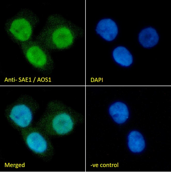
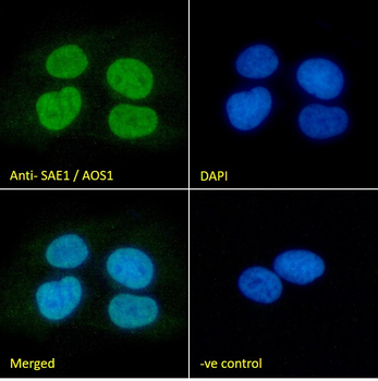
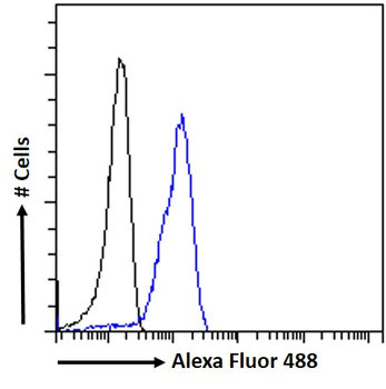
Goat anti-SAE1 / AOS1 Antibody [orb18563]
ELISA, FC, IF
Bovine, Canine, Human
Goat
Polyclonal
Unconjugated
100 μg - Item 1 of 6
Anti-ATG7 Antibody [orb654295]
FC, IHC, WB
Human, Mouse, Rat
Rabbit
Polyclonal
Unconjugated
100 μg, 10 μg - Item 1 of 6
- Item 1 of 4
APPBP1 Rabbit Polyclonal Antibody [orb155706]
IF, IHC-Fr, IHC-P, WB
Canine, Equine, Porcine, Rabbit
Human, Mouse, Rat
Rabbit
Polyclonal
Unconjugated
100 μl, 200 μl, 50 μl - Item 1 of 5
Anti-UBE1C/UBA3 Antibody [orb334570]
FC, ICC, IF, IHC, WB
Human, Mouse, Rat
Rabbit
Polyclonal
Unconjugated
10 μg, 100 μg