You have no items in your shopping cart.
Cart summary
Item 1 of 10
Item 1 of 10
TUBB3 (Neuronal Marker) Rabbit Polyclonal Antibody
Catalog Number: orb500814

Product Properties
| Catalog Number | orb500814 |
|---|---|
| Category | Antibodies |
| Description | TUBB3 (Neuronal Marker) Rabbit Polyclonal Antibody |
| Target | TUBB3 |
| Clonality | Polyclonal |
| Species/Host | Rabbit |
| Isotype | IgG |
| Conjugation | Unconjugated |
| Reactivity | Human, Mouse, Rat |
| Predicted Reactivity | Canine, Rabbit |
| Form/Appearance | Liquid |
| Concentration | 1mg/ml |
| Buffer/Preservatives | 0.01M TBS (pH7.4) with 1% rAlbumin, 0.02% Proclin300 and 50% Glycerol. |
| Purification | Affinity purified by Protein A |
| Immunogen | KLH conjugated synthetic peptide derived from human beta III Tubulin (401-450/450aa) |
| UniProt ID | Q13509 |
| MW | 50-55 kDa |
| Tested applications | FC, ICC, IF, IHC-Fr, IHC-P, WB |
| Dilution range | WB=1:500-2000, IHC-P=1:100-500, IHC-F=1:100-500, ICC/IF=1:100-500, IF=1:200-800, Flow-Cyt=1μg/Test |
| Antibody Type | Primary Antibody |
| Storage | Maintain refrigerated at 2-8°C for up to 2 weeks. For long term storage store at -20°C in small aliquots to prevent freeze-thaw cycles. |
| Alternative names | Neuron specific beta III Tubulin; beta 4; MC1R; TB Read more... |
| Research Area | Neurology, Neuroscience, Signal Transduction |
| Note | For research use only |
| Expiration Date | 12 months from date of receipt. |
Images

Blank control (blue): U-87MG Cells (fixed with 2% paraformaldehyde (10 min)). Primary Antibody: Rabbit Anti-MGLUR3/AF647 Conjugated antibody, dilution: 1 µg in 100 µl 1X PBS containing 0.5% BSA, Isotype Control Antibody: Rabbit IgG/AF647 (orange), used under the same conditions.
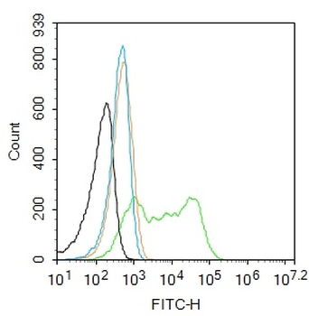
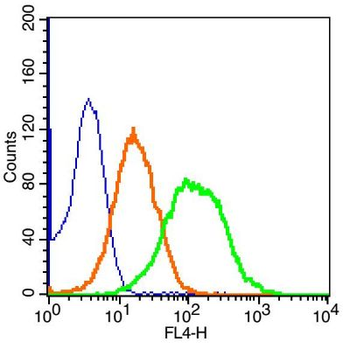
Blank control: SH-SY5Y. Primary Antibody (green line): Rabbit Anti-TUBB3 (Neuronal Marker) antibody (orb500814), dilution: 1 ug/Test, Secondary Antibody: Goat anti-rabbit IgG-FITC, dilution: 0.5 ug/Test. Protocol, The cells were fixed with 4% PFA (10 min at room temperature) and then permeabilized with 90% ice-cold methanol for 20 min at -20°C. The cells were then incubated in 5% BSA to block non-specific protein-protein interactions for 30 min at room temperature. Cells stained with Primary Antibody for 30 min at room temperature. The secondary antibody used for 40 min at room temperature. Acquisition of 20000 events was performed.
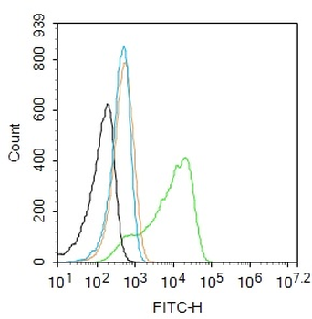
Blank control: SH-SY5Y. Primary Antibody (green line): Rabbit Anti-TUBB3 (Neuronal Marker) antibody (orb500814), dilution: 1 ug/Test, Secondary Antibody: Goat anti-rabbit IgG-FITC, dilution: 0.5 ug/Test. Protocol, The cells were fixed with 4% PFA (10 min at room temperature) and then permeabilized with 90% ice-cold methanol for 20 min at -20°C. The cells were then incubated in 5% BSA to block non-specific protein-protein interactions for 30 min at room temperature. Cells stained with Primary Antibody for 30 min at room temperature. The secondary antibody used for 40 min at room temperature. Acquisition of 20000 events was performed.
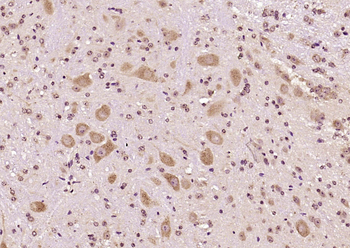
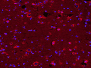
Paraformaldehyde-fixed, paraffin embedded (Mouse brain), Antigen retrieval by boiling in sodium citrate buffer (pH6.0) for 15 min, Block endogenous peroxidase by 3% hydrogen peroxide for 20 minutes, Blocking buffer (normal goat serum) at 37°C for 30 min, Antibody incubation with (TUBB3) Polyclonal Antibody, Unconjugated (orb500814) at 1:400 overnight at 4°C, followed by a conjugated Goat Anti-Rabbit IgG antibody for 90 minutes, and DAPI for nuclei staining.
Paraformaldehyde-fixed, paraffin embedded (mouse cerebellum), Antigen retrieval by boiling in sodium citrate buffer (pH6.0) for 15 min, Block endogenous peroxidase by 3% hydrogen peroxide for 20 minutes, Blocking buffer (normal goat serum) at 37°C for 30 min, Antibody incubation with (TUBB3 (Neuronal Marker)) Polyclonal Antibody, Unconjugated (orb500814) at 1:200 overnight at 4°C, followed by operating according to SP Kit (Rabbit) instructionsand DAB staining.
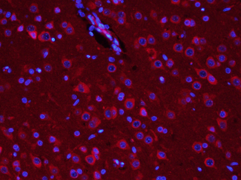
Paraformaldehyde-fixed, paraffin embedded (Rat brain), Antigen retrieval by boiling in sodium citrate buffer (pH6.0) for 15 min, Block endogenous peroxidase by 3% hydrogen peroxide for 20 minutes, Blocking buffer (normal goat serum) at 37°C for 30 min, Antibody incubation with (TUBB3) Polyclonal Antibody, Unconjugated (orb500814) at 1:400 overnight at 4°C, followed by a conjugated Goat Anti-Rabbit IgG antibody for 90 minutes, and DAPI for nuclei staining.
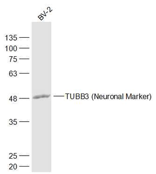
Sample: BV-2 (Rat) Cell Lysate at 30 ug, Primary: Anti-TUBB3 (Neuronal Marker) (orb500814) at 1/1000 dilution, Secondary: IRDye800CW Goat Anti-Rabbit IgG at 1/20000 dilution, Predicted band size: 50-55 kD, Observed band size: 50 kD.
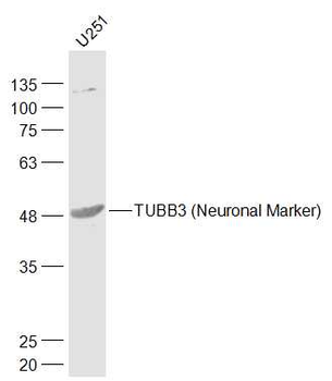
Sample: U251 (Human) Cell Lysate at 30 ug, Primary: Anti-TUBB3 (Neuronal Marker) (orb500814) at 1/1000 dilution, Secondary: IRDye800CW Goat Anti-Rabbit IgG at 1/20000 dilution, Predicted band size: 50-55 kD, Observed band size: 50 kD.
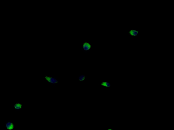
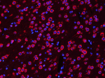
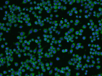
SH-SY5Y cell, 4% Paraformaldehyde-fixed, Ice-cold methanol at -20°C for 20 min, Blocking buffer (normal goat serum) at 37°C for 20 min, Antibody incubation with (TUBB3) polyclonal Antibody, Unconjugated (orb500814) 1:100, 90 minutes at 37°C, followed by a FITC conjugated Goat Anti-Rabbit IgG antibody at 37°C for 90 minutes, DAPI (blue) was used to stain the cell nuclei.
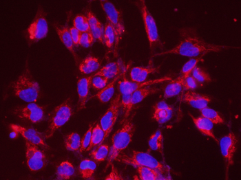
Tissue/Cell: BV-2 cell, 4% Paraformaldehyde-fixed, Triton X-100 at room temperature for 20 min, Blocking buffer (normal goat serum) at 37°C for 20 min, Antibody incubation with (TUBB3) Polyclonal Antibody, Unconjugated (orb500814) 1:200, 90 minutes at 37°C, followed by a conjugated Goat Anti-Rabbit IgG antibody (orb868805) at 37°C for 90 minutes, DAPI (5 ug/ml, blue) was used to stain the cell nuclei.
Similar Products
- Item 1 of 2
TUBB3 (Neuronal Marker) Rabbit Polyclonal Antibody (BF594) [orb1587539]
FC, ICC, IF
Canine, Rabbit
Human, Mouse, Rat
Rabbit
Polyclonal
BF594
100 μl - Item 1 of 1
TUBB3 (Neuronal Marker) Rabbit Polyclonal Antibody (BF647) [orb1587538]
FC, ICC, IF
Canine, Rabbit
Human, Mouse, Rat
Rabbit
Polyclonal
BF647
100 μl 
TUBB3 (Neuronal Marker) Rabbit Polyclonal Antibody (PE) [orb484855]
FC, ICC, IF
Canine, Rabbit
Human, Mouse, Rat
Rabbit
Polyclonal
PE
TUBB3 (Neuronal Marker) Rabbit Polyclonal Antibody (FITC) [orb466084]
FC, ICC, IF
Canine, Rabbit
Human, Mouse, Rat
Rabbit
Polyclonal
FITC
100 μlTUBB3 (Neuronal Marker) Rabbit Polyclonal Antibody (HRP) [orb468976]
IHC-Fr, IHC-P, WB
Canine, Rabbit
Human, Mouse, Rat
Rabbit
Polyclonal
HRP
100 μl
Reviews
TUBB3 (Neuronal Marker) Rabbit Polyclonal Antibody (orb500814)

- 0.0
Based on 0 reviews
Participating in our Biorbyt product reviews program enables you to support fellow scientists by sharing your firsthand experience with our products.
Login to Submit a Review