You have no items in your shopping cart.
Cart summary
Item 1 of 5
Item 1 of 5
TPI1 Antibody
Catalog Number: orb1264514

| Catalog Number | orb1264514 |
|---|---|
| Category | Antibodies |
| Description | TPI1 Antibody |
| Species/Host | Rabbit |
| Clonality | Polyclonal |
| Tested applications | FC, IHC-P, WB |
| Predicted Reactivity | Bovine, Monkey, Porcine, Rabbit, Rat |
| Reactivity | Human, Mouse |
| Isotype | Rabbit Ig |
| Immunogen | This TPI1 antibody is generated from rabbits immunized with a KLH conjugated synthetic peptide between 68-96 amino acids from the N-terminal region of human TPI1. |
| Antibody Type | Primary Antibody |
| Concentration | batch dependent |
| Form/Appearance | Liquid |
| Conjugation | Unconjugated |
| MW | 31 kDa |
| Target | TPI1 |
| UniProt ID | P60174 |
| NCBI | P60174 |
| Storage | Maintain refrigerated at 2-8°C for up to 2 weeks. For long term storage store at -20°C in small aliquots to prevent freeze-thaw cycles. |
| Buffer/Preservatives | Supplied in PBS with 0.09% (W/V) sodium azide. |
| Alternative names | Triosephosphate isomerase, TIM, Triose-phosphate i Read more... |
| Note | For research use only |
| Application notes | For WB starting dilution is: 1:1000For IHC-P starting dilution is: 1:10~50For FACS starting dilution is: 1:10~50 |
| Expiration Date | 12 months from date of receipt. |
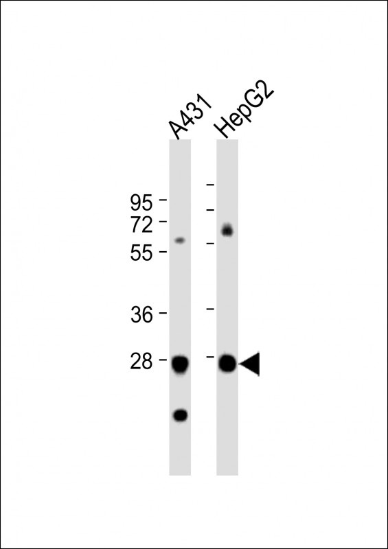
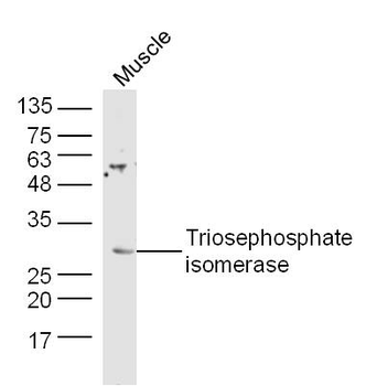
Western Blot at 1:1000 dilution Lane 1: A431 whole cell lysate Lane 2: HepG2 whole cell lysate Lysates/proteins at 20 ug per lane.
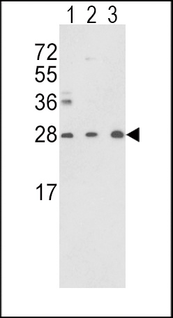
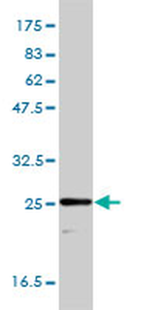
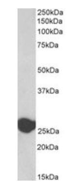
Western blot analysis of TPI1 Antibody in Y79 (lane 1), CEM (lane 2) cell line and mouse brain tissue (lane 3) lysates (35 ug/lane) (2ug/ml)
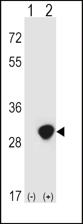
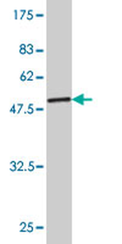
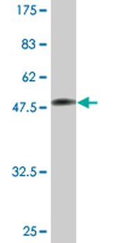
Western blot analysis of TPI1 using rabbit polyclonal TPI1 Antibody using 293 cell lysates (2 ug/lane) either nontransfected (Lane 1) or transiently transfected (Lane 2) with the TPI1 gene.

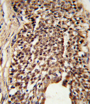
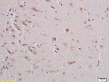
Formalin-fixed and paraffin-embedded human prostate carcinoma reacted with TPI1 Antibody (N-term), which was peroxidase-conjugated to the secondary antibody, followed by DAB staining.
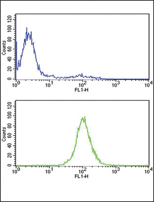
Flow cytometric analysis of CEM cells (bottom histogram) compared to a negative control cell (top histogram). FITC-conjugated goat-anti-rabbit secondary antibodies were used for the analysis.
- Item 1 of 5
TPI1 Antibody (N-term) [orb1931341]
FC, IHC-P, WB
Porcine, Rabbit, Rat
Human, Mouse
Rabbit
Polyclonal
Unconjugated
100 μl, 50 μl - Item 1 of 3
TPI1 monoclonal antibody (M01), clone 1D10-2E2 [orb2292033]
ELISA, WB
Human
Mouse
Monoclonal
Unconjugated
100 μg - Item 1 of 3
Triosephosphate isomerase Rabbit Polyclonal Antibody [orb7113]
IF, IHC-Fr, IHC-P, WB
Bovine, Canine, Equine, Gallus, Porcine, Rabbit, Sheep
Human, Mouse, Rat
Rabbit
Polyclonal
Unconjugated
100 μl, 200 μl, 50 μl - Item 1 of 1
Goat anti-Triosephosphate isomerase, Biotinylated Antibody [orb389379]
ELISA, IHC, WB
Bovine, Canine, Human, Mouse, Rat
Goat
Polyclonal
Unconjugated
100 μg - Item 1 of 2
TPI1 monoclonal antibody (M02A), clone 2C3 [orb2292032]
ELISA, WB
Human
Mouse
Monoclonal
Unconjugated
200 μl