You have no items in your shopping cart.
Cart summary
Item 1 of 7
Item 1 of 7
TIGIT Antibody
Catalog Number: orb1240137
| Catalog Number | orb1240137 |
|---|---|
| Category | Antibodies |
| Description | TIGIT Antibody |
| Species/Host | Mouse |
| Clonality | Monoclonal |
| Clone Number | 4A10 |
| Tested applications | ELISA, FC, ICC, IF, IHC-P, WB |
| Reactivity | Human |
| Isotype | IgG1 |
| Immunogen | TIGIT antibody was raised against the extracellular domain of human TIGIT |
| Concentration | 1 mg/mL |
| Dilution range | TIGIT antibody can be used for immunohistochemistry starting at 2 μg/mL. For immunofluorescence start at 1 μg/mL. For flow cytometry at 1 μg/ml. For immunocytochemistry at 1 μg/mL. For Western blot at 1 μg/mL. Antibody validated: Western Blot in human samples; Immunohistochemistry in human samples; Immunocytochemistry in human samples; Immunofluorescence in human samples and Flow Cytometry in human samples. All other applications and species not yet tested. |
| Form/Appearance | Liquid |
| Conjugation | Unconjugated |
| MW | Predicted: 26 kDa Observed: 47 kDa |
| Target | TIGIT |
| UniProt ID | Q495A1 |
| NCBI | NP_776160 |
| Storage | TIGIT antibody can be stored at 4°C for three months and -20°C, stable for up to one year. As with all antibodies care should be taken to avoid repeated freeze thaw cycles. Antibodies should not be exposed to prolonged high temperatures. |
| Buffer/Preservatives | TIGIT Antibody is supplied in PBS containing 0.02% sodium azide and 50% glycerol. |
| Alternative names | TIGIT Antibody: T-cell immunoreceptor with Ig and Read more... |
| Note | For research use only |
| Application notes | TIGIT antibody can be used for immunohistochemistry starting at 2 μg/mL. For immunofluorescence start at 1 μg/mL. For flow cytometry at 1 μg/ml. For immunocytochemistry at 1 μg/mL. For Western blot at 1 μg/mL. Antibody validated: Western Blot in human samples; Immunohistochemistry in human samples; Immunocytochemistry in human samples; Immunofluorescence in human samples and Flow Cytometry in human samples. All other applications and species not yet tested. |
| Expiration Date | 12 months from date of receipt. |
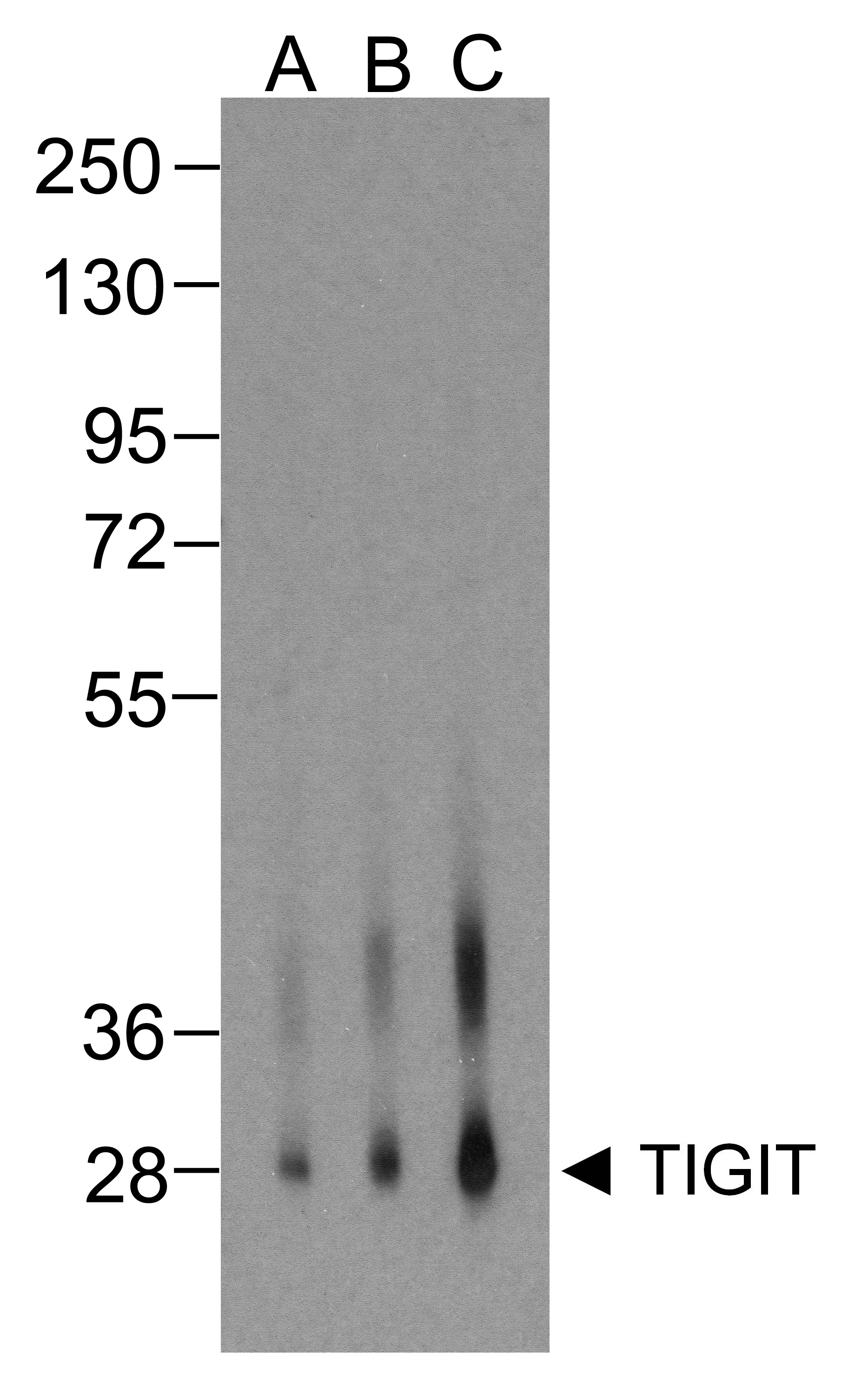
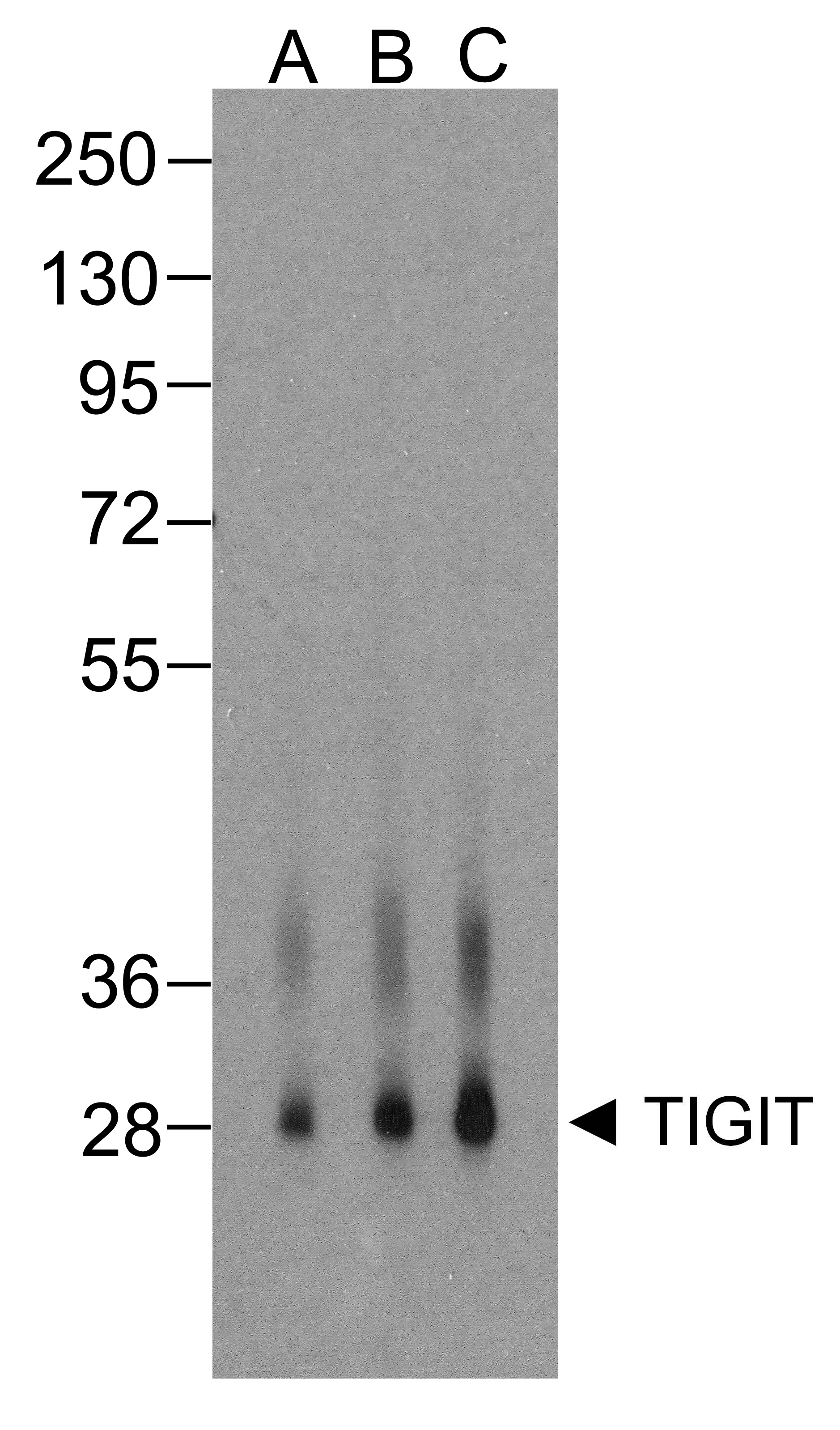
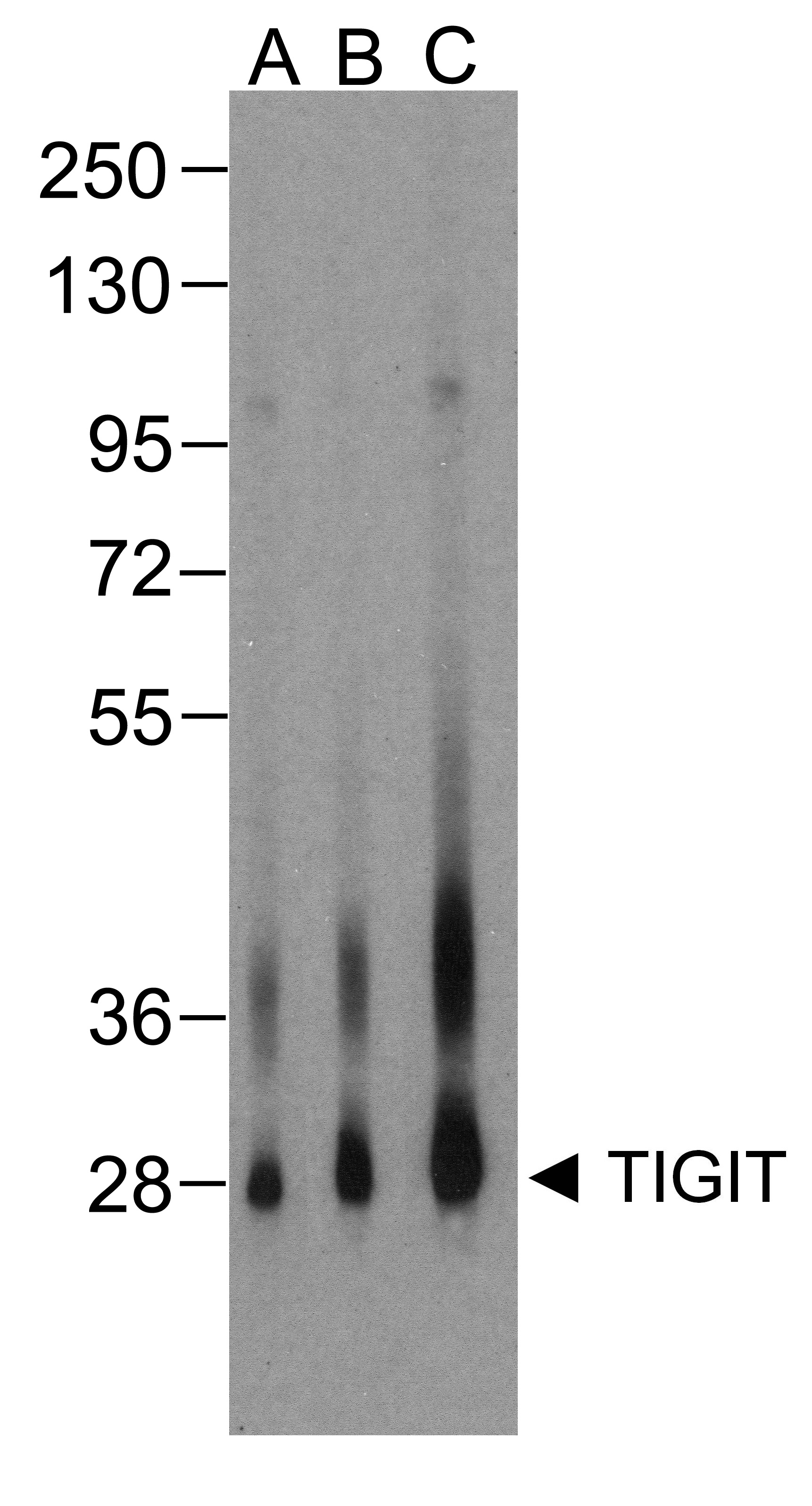
Western blot analysis of TIGIT in over expressing HEK293 cells using orb1240137 antibody at (A) 0.25 μg/ml, (B) 0.5 μg/ml, and (C) 1 μg/ml.
Immunocytochemistry of TIGIT in over expressing HEK293 cells using TIGIT antibody and control mouse IgG antibody (left corner box) at 1 μg/ml.
Immunofluorescence of TIGIT in over expressing HEK293 cells using TIGIT Antibody at 1 μg/ml. Green: TIGIT Antibody [4A10] (orb1240137) Blue: DAPI staining
Immunofluorescence of TIGIT in human stomach carcinoma tissue using TIGIT Antibody at 5 μg/ml. Green: TIGIT Antibody [4A10] (orb1240137) Blue: DAPI staining
Immunohistochemistry of TIGIT in human stomach carcinoma tissue using TIGIT Antibody and control mouse IgG (corner box) at 2 μg/ml.
Flow cytometry analysis of TIGIT over expressing HEK293 cells using TIGIT antibody at 1 μg/ml. Blue: untransfected HEK293 cells. Yellow: TIGIT over expressing HEK293 cells.

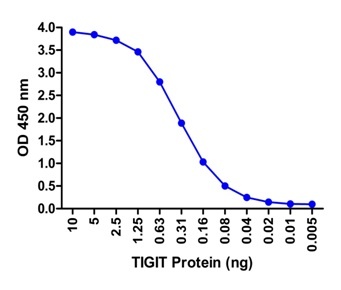
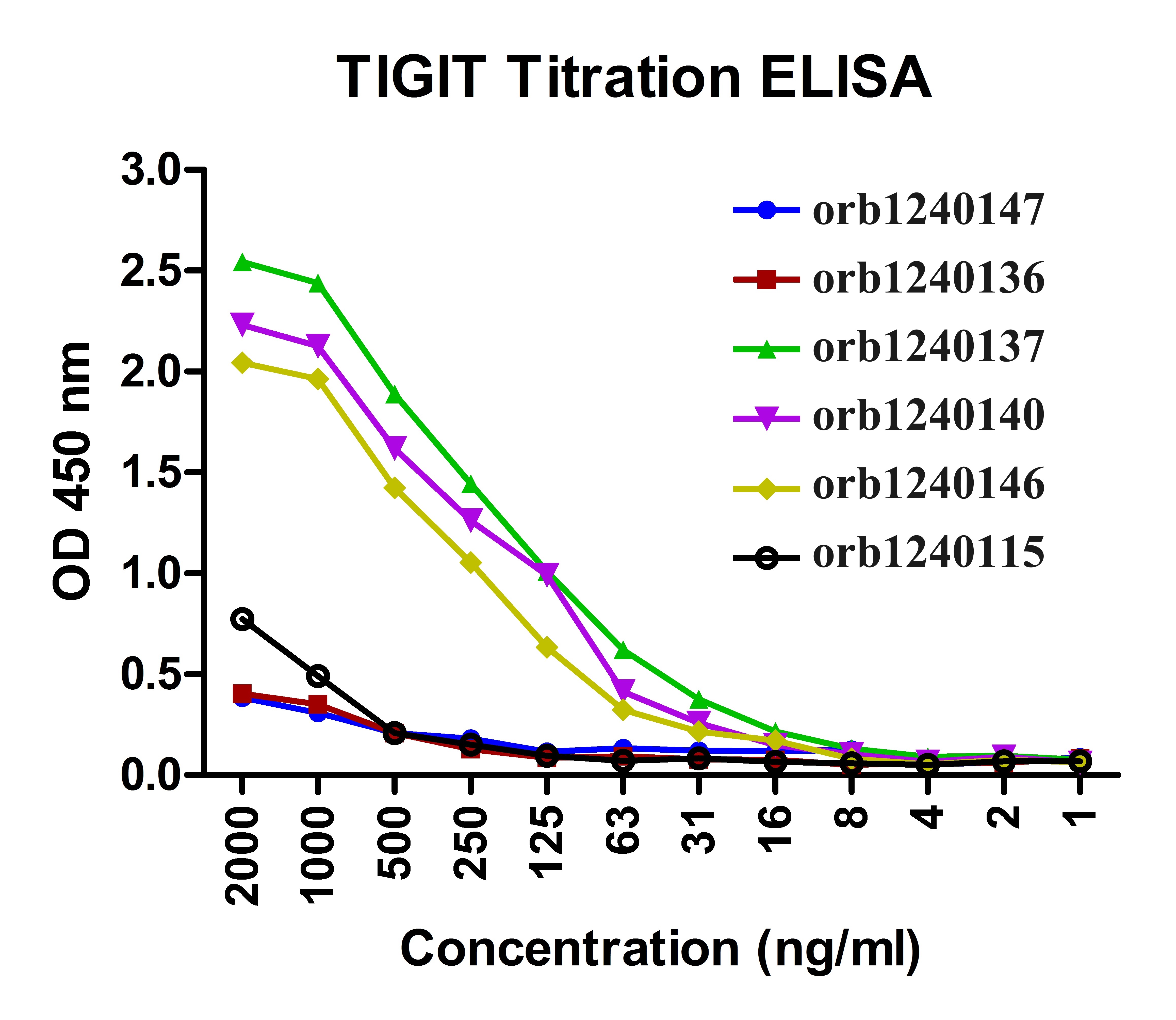
Titration curve analysis of TIGIT mAbs to detect recombinant TIGIT in ELISA with orb1240147, orb1240136, orb1240137, orb1240140, orb1240146 and orb1240115 antibodies at decreasing concentrations.
- Item 1 of 8
- Item 1 of 7
- Item 1 of 6
- Item 1 of 6
- Item 1 of 6


Submit a review
Filter by Rating

- 5 stars

- 4 stars
- 3 stars
- 2 stars
- 1 stars