You have no items in your shopping cart.
Cart summary
Item 1 of 3
Item 1 of 3
TGFB3 Antibody / TGF beta 3
Catalog Number: orb1825518

| Catalog Number | orb1825518 |
|---|---|
| Category | Antibodies |
| Description | Transforming growth factor betas (TGF-betas) were originally discovered due to their ability to promote anchorage-independent growth of rat NRK fibroblasts in the presence of TGF-beta. TGF-beta 1, TGF-beta 2 and TGF-beta 3 are each synthesized as precursor proteins that are very similar in that each is cleaved to yield a 112 amino acid polypeptide that remains associated with the latent portion of the molecules. TGF-beta 3 mediates many intercellular interactions that occur during embryonic development, cell differentiation and epithelial homeostasis. TGF-beta 3 overexpresses in extramammary Paget s disease (EPD) and downregulates in Bowen s disease, indicating that its expression is a useful indicator of tumor activity. TGF-beta 3 levels strongly correlate with IGF-1 and osteocalcin levels in serum. Significant amounts of TGF-beta 3 circulation appear to be representative of TGFbeta 3 expression in bone and may in part be derived from bone. Glucocorticoids may block TGF-beta production by modulating mRNA levels and c-Jun activity. |
| Clonality | Monoclonal |
| Species/Host | Mouse |
| Isotype | Mouse IgG2, kappa |
| Conjugation | Unconjugated |
| Reactivity | Human |
| Immunogen | A recombinant partial protein sequence (within amino acids 50-250) from the human protein was used as the immunogen for the TGF beta 3 antibody. |
| UniProt ID | P10600 |
| Tested applications | IHC-P |
| Dilution range | Immunohistochemistry (FFPE): 1-2ug/ml for 30 minutes at RT |
| Antibody Type | Primary Antibody |
| Clone Number | TGFB3/4801 |
| Formula | 0.2 mg/ml in 1X PBS with 0.1 mg/ml BSA (US sourced), 0.05% sodium azide |
| Storage | Maintain refrigerated at 2-8°C for up to 2 weeks. For long term storage store at -20°C in small aliquots to prevent freeze-thaw cycles. |
| Hazard Information | This TGF beta 3 antibody is available for research use only. |
| Note | For research use only |

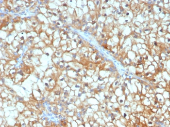
IHC staining of FFPE human renal cell carcinoma tissue with TGF beta 3 antibody (clone TGFB3/4801). HIER: boil tissue sections in pH9 10 mM Tris with 1 mM EDTA for 20 min and allow to cool before testing.
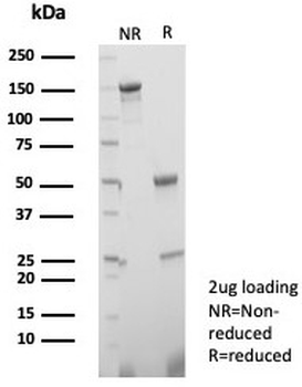
SDS-PAGE analysis of purified, BSA-free TGF beta 3 antibody (clone TGFB3/4801) as confirmation of integrity and purity.
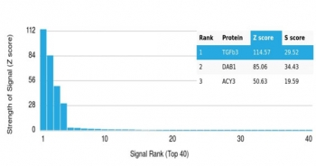
Analysis of a HuProt (TM) microarray containing more than 19000 full-length human proteins using TGF beta 3 antibody (clone TGFB3/4801). Z- and S- Score: The Z-score represents the strength of a signal that a monoclonal antibody (in combination with a fluorescently-tagged anti-IgG secondary antibody) produces when binding to a particular protein on the HuProt (TM) array. Z-scores are described in units of standard deviations (SD's) above the mean value of all signals generated on that array. If targets on HuProt (TM) are arranged in descending order of the Z-score, the S-score is the difference (also in units of SD's) between the Z-score. S-score therefore represents the relative target specificity of a mAb to its intended target. A mAb is considered to specific to its intended target, if the mAb has an S-score of at least 2.5. For example, if a mAb binds to protein X with a Z-score of 43 and to protein Y with a Z-score of 14, then the S-score for the binding of that mAb to protein X is equal to 29.
- Item 1 of 3