You have no items in your shopping cart.
Cart summary
Item 1 of 5
Item 1 of 5
TBX1 Antibody (C-term)
Catalog Number: orb1927949

| Catalog Number | orb1927949 |
|---|---|
| Category | Antibodies |
| Description | Affinity Purified Rabbit Polyclonal Antibody (Pab) |
| Target | This TBX1 antibody is generated from rabbits immunized with a KLH conjugated synthetic peptide between 327-356 amino acids from the C-terminal region of human TBX1. |
| Clonality | Polyclonal |
| Species/Host | Rabbit |
| Isotype | Rabbit IgG |
| Conjugation | Unconjugated |
| Reactivity | Human |
| Form/Appearance | Purified polyclonal antibody supplied in PBS with 0.09% (W/V) sodium azide. This antibody is purified through a protein A column, followed by peptide affinity purification. |
| UniProt ID | O43435 |
| MW | 43133 Da |
| Tested applications | IF, IHC-P, WB |
| Dilution range | IF: 1:10~50, WB: 1:1000, WB: 1:1000, IHC-P: 1:50~100, IHC-P: 1:25 |
| Clone Number | RB22154 |
| Storage | Maintain refrigerated at 2-8°C for up to 2 weeks. For long term storage store at -20°C in small aliquots to prevent freeze-thaw cycles |
| Alternative names | T-box transcription factor TBX1, T-box protein 1, Read more... |
| Note | For research use only |
| NCBI | NP_542378.1, NP_005983.1, NP_542377.1 |
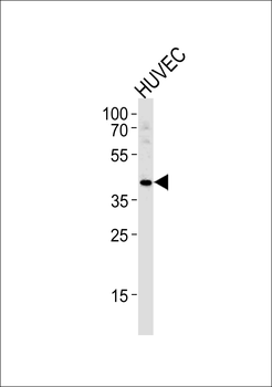
TBX1 Antibody (C-term) western blot analysis in HLVEC cell line lysates (35 ug/lane).This demonstrates the TBX1 antibody detected the TBX1 protein (arrow).
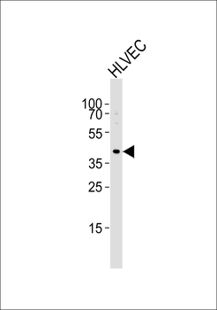
Western blot analysis of lysate from HLVEC cell line, using TBX1 Antibody (C-term). Diluted at 1:1000 at each lane. A goat anti-rabbit IgG H&L (HRP) at 1:5000 dilution was used as the secondary antibody. Lysate at 35 ug.
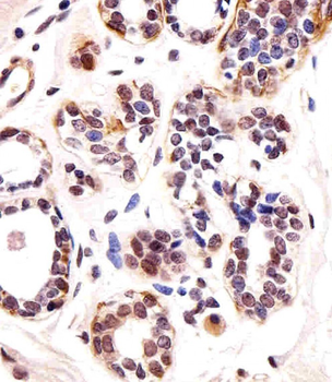
Immunohistochemical analysis of paraffin-embedded H. breast section using TBX1 Antibody (C-term). Diluted at 1:25 dilution. A undiluted biotinylated goat polyvalent antibody was used as the secondary, followed by DAB staining.

Formalin-fixed and paraffin-embedded human testis tissue reacted with TBX1 Antibody (C-term), which was peroxidase-conjugated to the secondary antibody, followed by DAB staining. This data demonstrates the use of this antibody for immunohistochemistry; clinical relevance has not been evaluated.
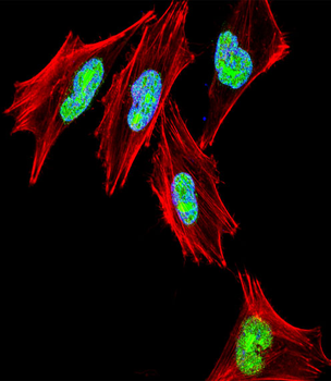
Fluorescent confocal image of A2058 cell stained with TBX1 Antibody (C-term). A2058 cells were fixed with 4% PFA (20 min), permeabilized with Triton X-100 (0.1%, 10 min), then incubated with TBX1 primary antibody (1: 25, 1 h at 37°C). For secondary antibody, Alexa Fluor 488 conjugated donkey anti-rabbit antibody (green) was used (1:400, 50 min at 37°C).Cytoplasmic actin was counterstained with Alexa Fluor 555 (red) conjugated Phalloidin (7units/ml, 1 h at 37°C). Nuclei were counterstained with DAPI (blue) (10 µg/ml, 10 min). TBX1 immunoreactivity is localized to nucleus significantly.
