You have no items in your shopping cart.
Cart summary
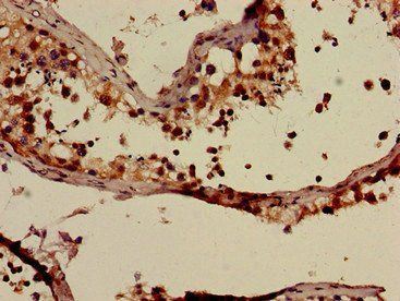
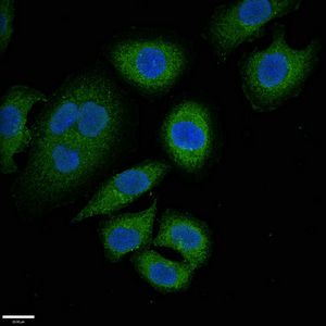
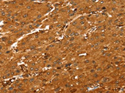
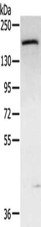
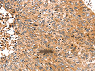
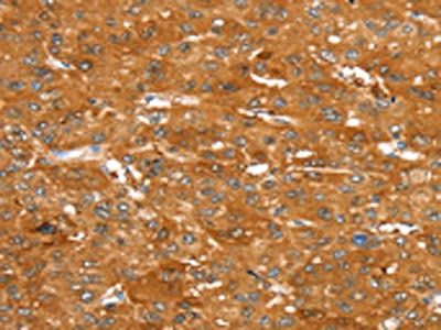
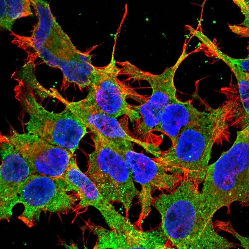
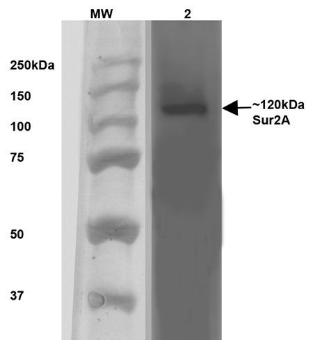
SUR2A antibody
Catalog Number: orb150354
| Catalog Number | orb150354 |
|---|---|
| Category | Antibodies |
| Description | Mouse monoclonal to SUR2A (Biotin). Sulfonylurea receptors (SUR) are membrane proteins which are the molecular targets of the sulfonylurea class of anti-diabetic drugs whose mechanism of action is to promote insulin release from pancreatic beta cells. More specifically, SUR proteins are subunits of the inward-rectifier potassium ion channels Kir6. x (6. 1 and 6. 2). The association of four K ir6. x and four SUR subunits form an ion conducting channel commonly referred to as the K ATP channel. The primary function of the sulfonylurea receptor is to sense intracellular levels of the nucleotides ATP and ADP and in response facilitate the open or closing its associated Kir 6. x potassium channel. Hence the K ATP channel monitors the energy balance within the cell.. |
| Species/Host | Mouse |
| Clonality | Monoclonal |
| Clone Number | S319A-14 |
| Tested applications | ICC, IHC, WB |
| Reactivity | Human, Mouse, Rat |
| Isotype | IgG2A |
| Immunogen | Fusion protein amino acids 1505-1546 (SSIVDAGLVLVFSEGILVECDTGPNLLQHKNGLFSTLVMTNK, cytoplasmic C-terminus) of mouse SUR2A |
| Concentration | 1 mg/ml |
| Dilution range | WB (1:1000) |
| Conjugation | Biotin |
| MW | 120kDa |
| Target | SUR2A |
| Entrez | 20928 |
| UniProt ID | P70170 |
| NCBI | NP_001038185.1 |
| Storage | Conjugated antibodies should be stored at 4°C |
| Buffer/Preservatives | PBS pH 7.4, 50% glycerol, 0.1% sodium azide *Storage buffer may change when conjugated |
| Alternative names | ABCC9 antibody, Sulfonylurea receptor 2 antibody, Read more... |
| Note | For research use only |
| Application notes | 1 µg/ml of SMC-431 was sufficient for detection of SUR2A in 20 µg of mouse brain membrane lysate and assayed by colorimetric immunoblot analysis using goat anti-mouse IgG:HRP as the secondary antibody. |
| Expiration Date | 12 months from date of receipt. |
- Item 1 of 2
- Item 1 of 2
ABCC9 antibody [orb525139]
ELISA, IHC, WB
Human, Mouse, Rat
Rabbit
Polyclonal
Unconjugated
50 μl, 100 μl - Item 1 of 2
- Item 1 of 2

Submit a review
Filter by Rating

- 5 stars

- 4 stars
- 3 stars
- 2 stars
- 1 stars