You have no items in your shopping cart.
Cart summary
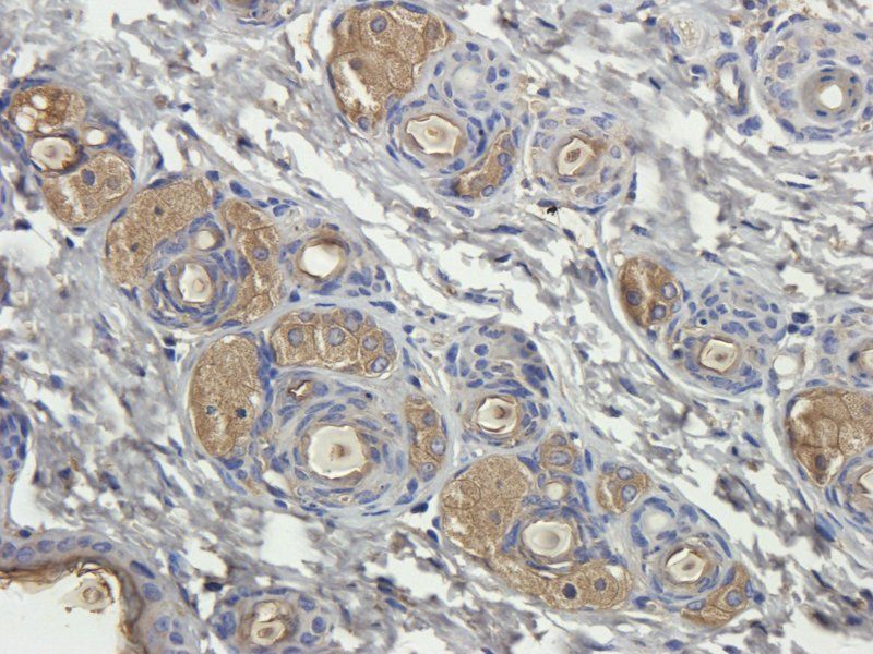
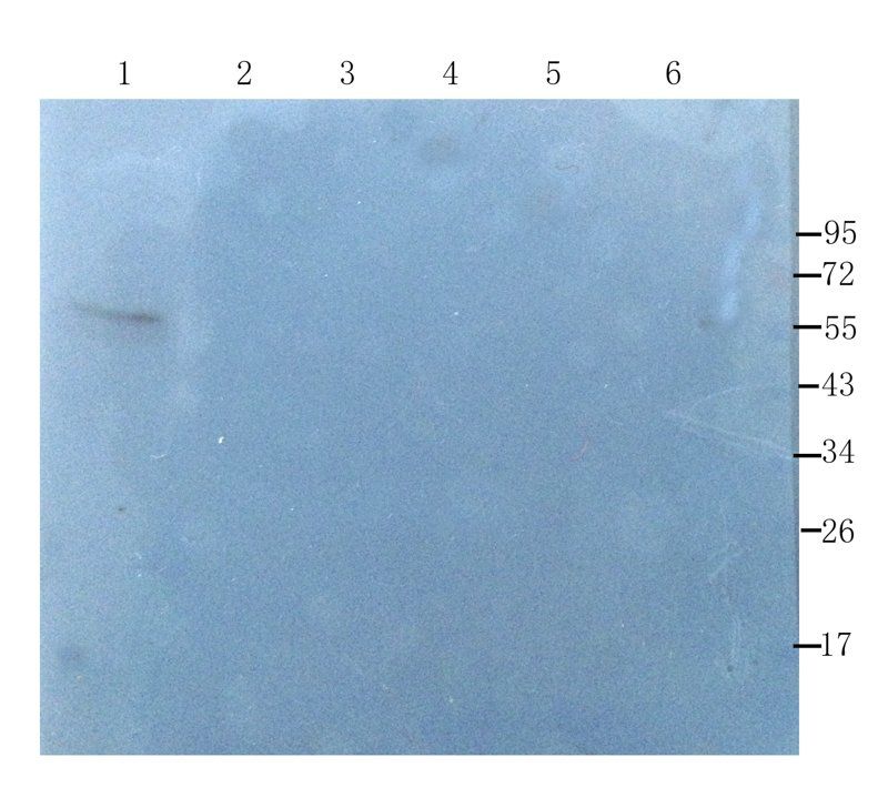
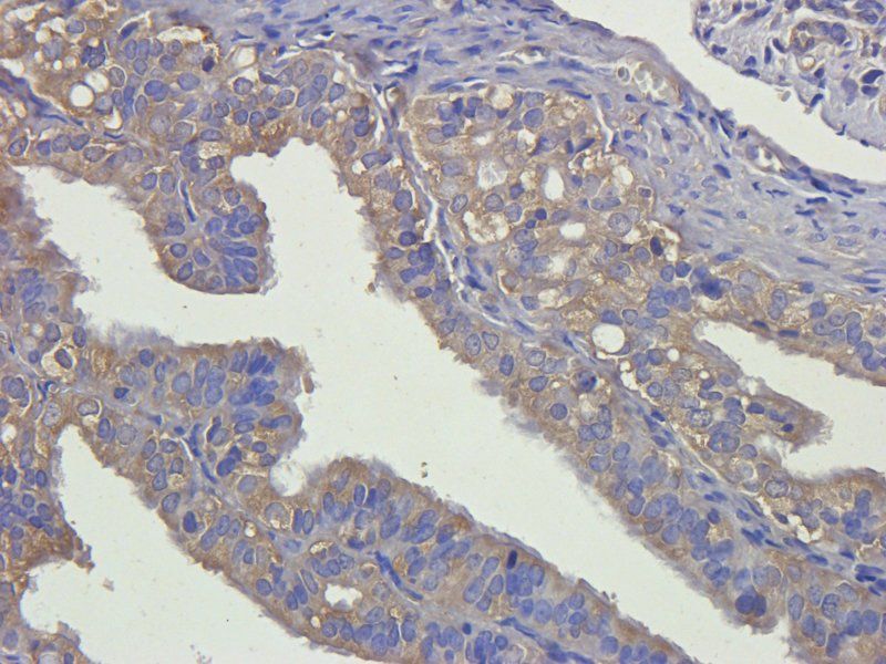
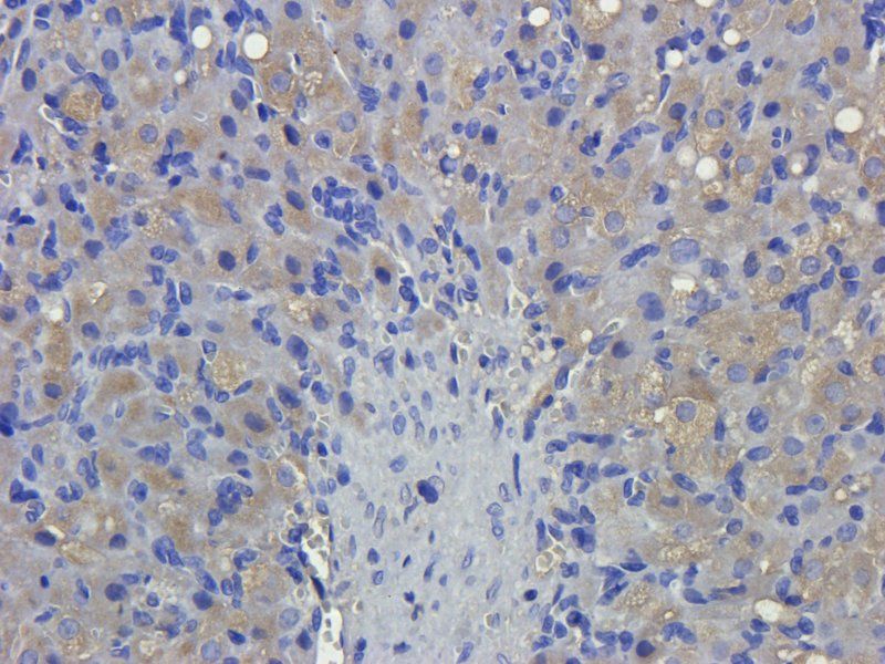
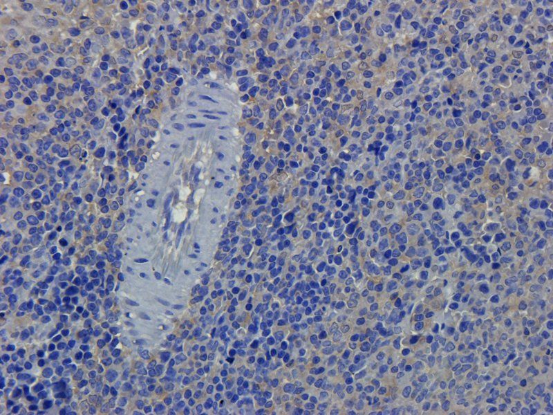
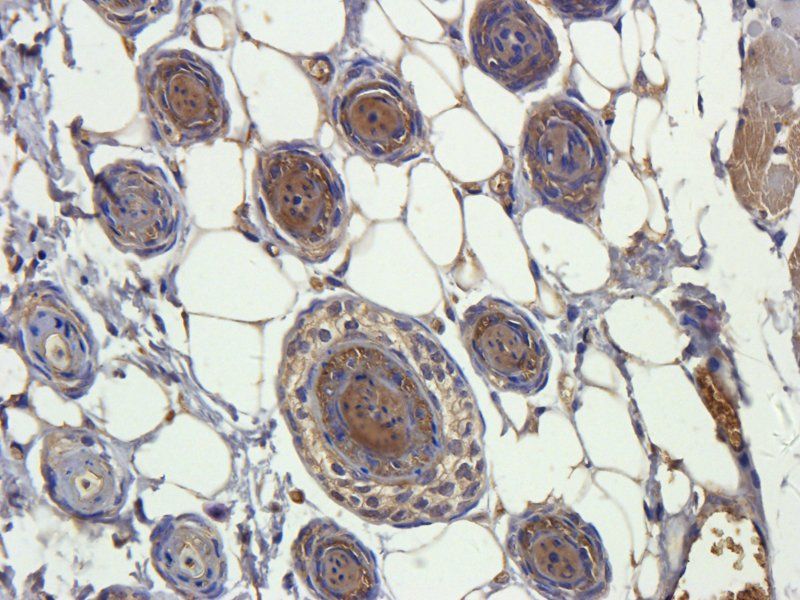
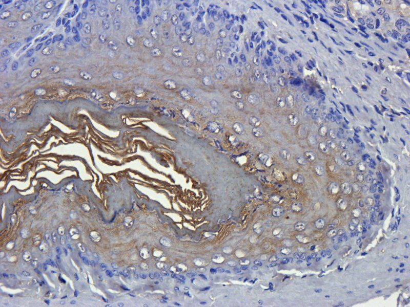
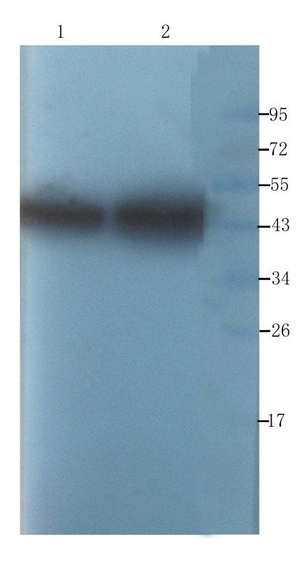
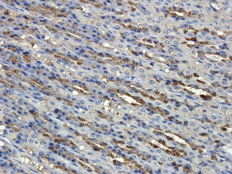
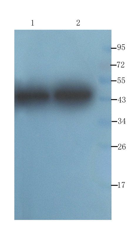
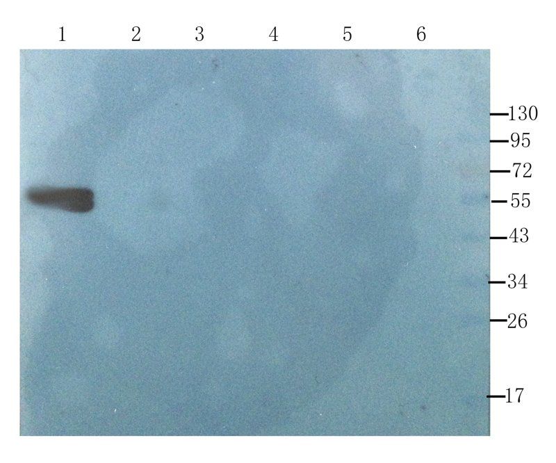
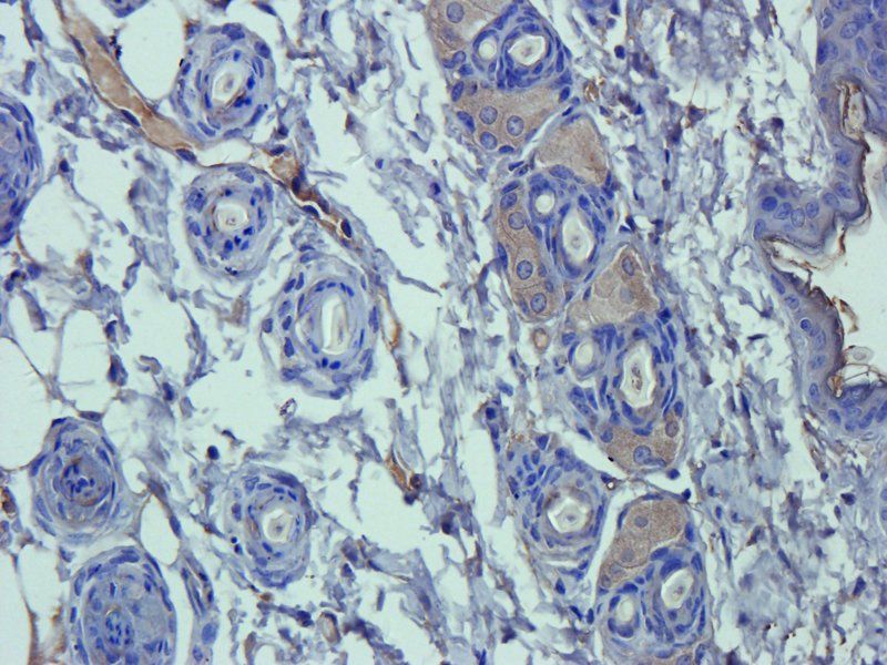
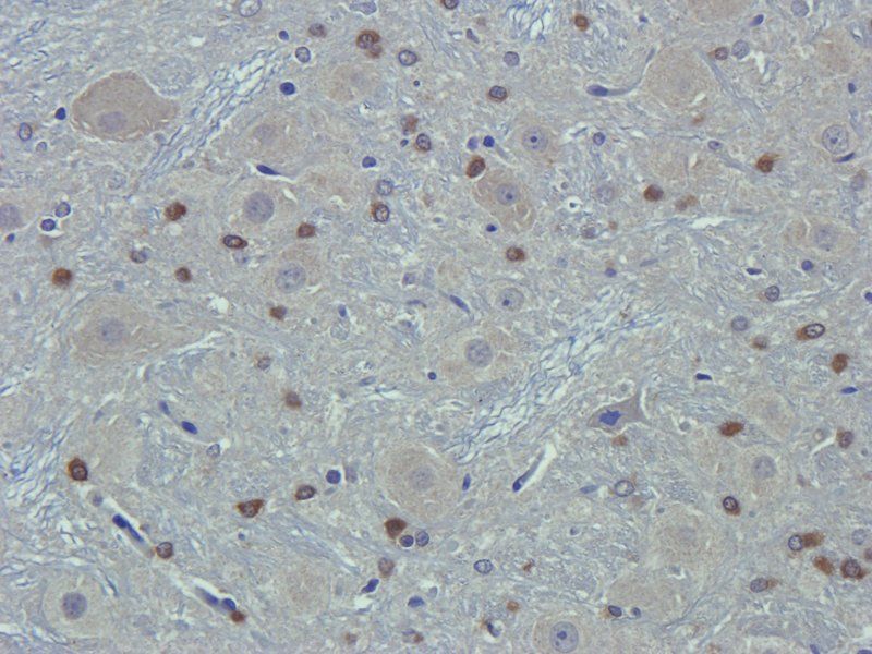
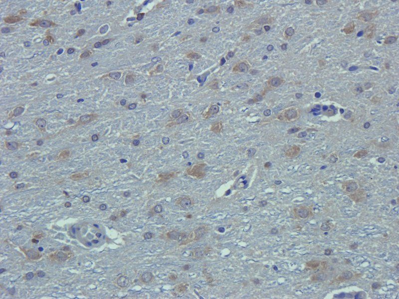
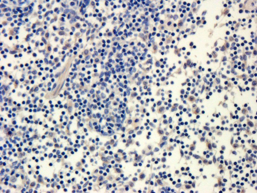
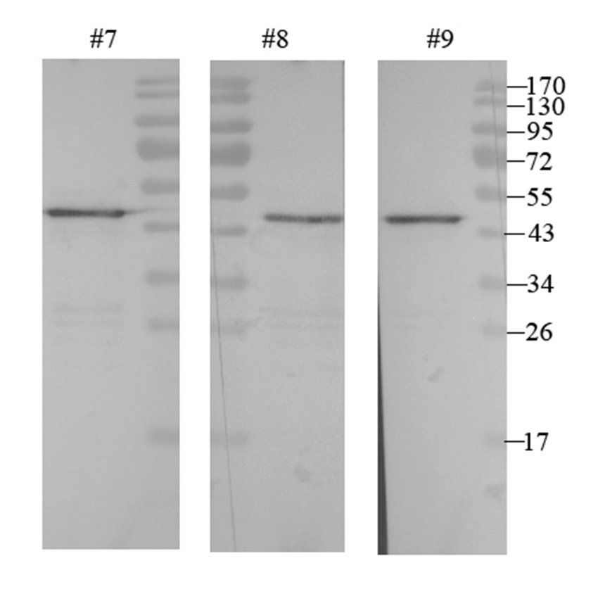
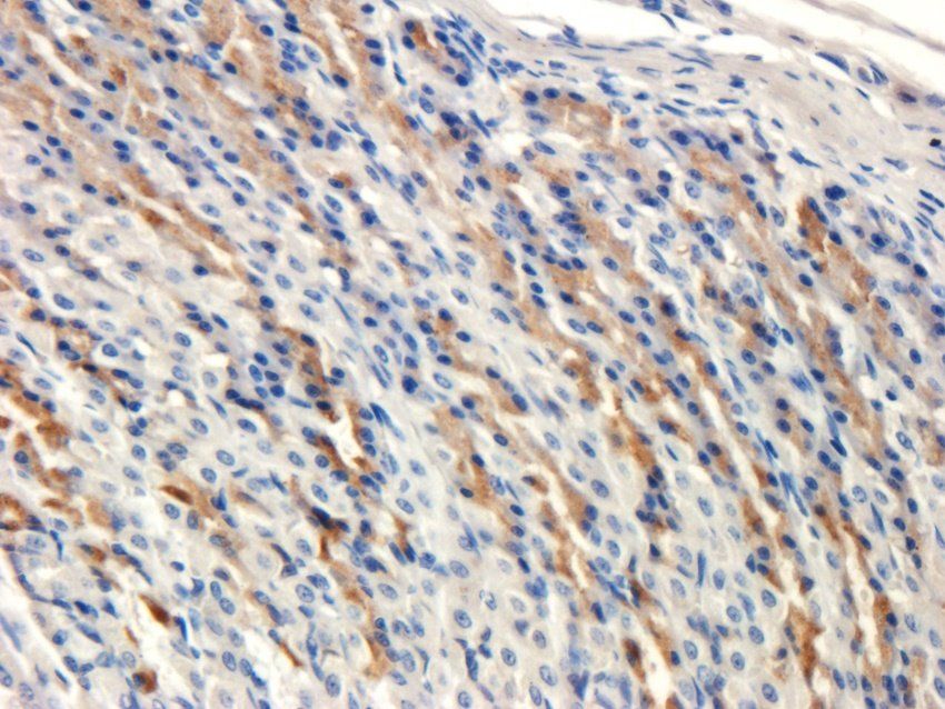
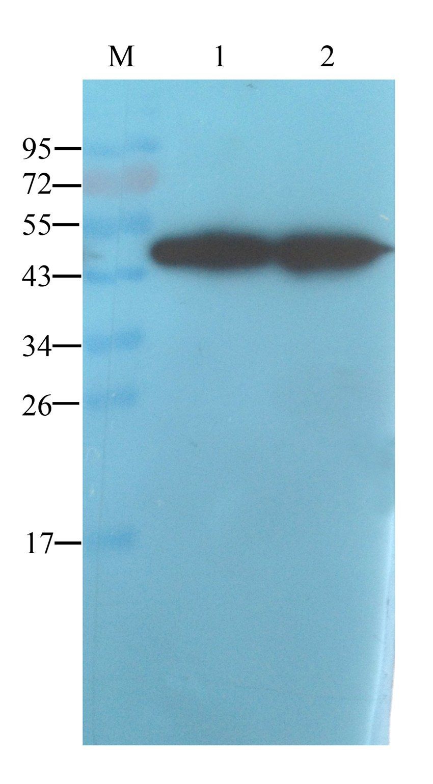
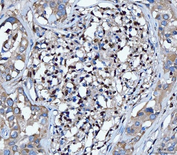
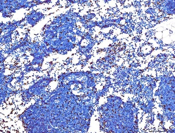
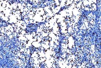
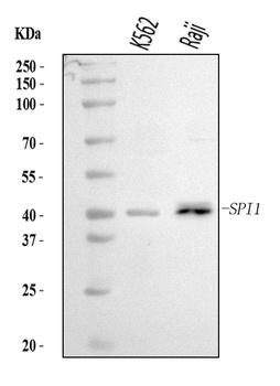
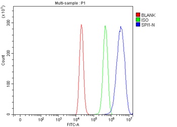
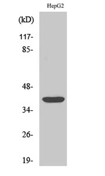
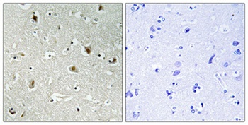
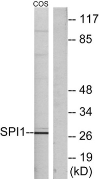
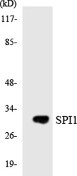
Spi1 antibody
Catalog Number: orb312530
| Catalog Number | orb312530 |
|---|---|
| Category | Tools |
| Description | Spi1 antibody |
| Species/Host | Rabbit |
| Clonality | Polyclonal |
| Tested applications | WB |
| Predicted Reactivity | Bovine, Equine, Mouse, Porcine, Rat, Sheep |
| Reactivity | Human |
| Isotype | IgG |
| Immunogen | KLH conjugated synthetic peptide derived from human PU.1/Spi1 (151-250/270 aa) |
| Concentration | 1mg/ml |
| Dilution range | WB=1:500-2000 |
| Form/Appearance | Liquid |
| Conjugation | Unconjugated |
| MW | 31 kDa |
| Target | PU.1/Spi1 |
| UniProt ID | P17947 |
| Storage | Shipped at 4°C. Store at -20 °C for one year. Avoid repeated freeze/thaw cycles. |
| Buffer/Preservatives | 0.01M TBS(pH7.4) with 1% rAlbumin, 0.03% Proclin300 and 50% Glycerol. |
| Alternative names | 31 kDa Transforming Protein; 31 kDa-transforming p Read more... |
| Note | For research use only |
| Expiration Date | 12 months from date of receipt. |
- Item 1 of 9
alpha 1 Antitrypsin antibody [orb339609]
IHC-P, WB
Mouse, Rat
Rabbit
Polyclonal
Unconjugated
100 μg, 200 μg - Item 1 of 6
alpha 1 Antitrypsin antibody [orb339610]
IHC-P, WB
Mouse, Rat
Rabbit
Polyclonal
Unconjugated
100 μg, 200 μg, 500 μg - Item 1 of 4
alpha 1 Antitrypsin antibody [orb334222]
ELISA, IHC-P, WB
Human, Rat
Mouse
Monoclonal
Unconjugated
100 μg, 200 μg - Item 1 of 5
PU.1/SPI1 Antibody [orb1097960]
ELISA, FC, IHC, WB
Human, Mouse, Rat
Rabbit
Polyclonal
Unconjugated
10 μg, 100 μg - Item 1 of 4
PU.1 antibody [orb766159]
ELISA, IF, IHC-P, WB
Human, Monkey, Mouse, Rat
Rabbit
Polyclonal
Unconjugated
50ul, 100ul

Submit a review
Filter by Rating

- 5 stars

- 4 stars
- 3 stars
- 2 stars
- 1 stars