You have no items in your shopping cart.
Cart summary
Item 1 of 5
Item 1 of 5
SP1 Antibody
Catalog Number: orb1270161
| Catalog Number | orb1270161 |
|---|---|
| Category | Antibodies |
| Description | SP1 Antibody |
| Species/Host | Rabbit |
| Clonality | Polyclonal |
| Tested applications | FC, IF, IHC-P, WB |
| Predicted Reactivity | Mouse, Rat |
| Reactivity | Human |
| Isotype | Rabbit Ig |
| Immunogen | This SP1 antibody is generated from rabbits immunized with a KLH conjugated synthetic peptide between 677-707 amino acids from the C-terminal region of human SP1. |
| Concentration | batch dependent |
| Dilution range | For WB starting dilution is: 1:500For IHC-P starting dilution is: 1:50~100For FACS starting dilution is: 1:10~50For IF starting dilution is: 1:10~50 |
| Form/Appearance | Liquid |
| Conjugation | Unconjugated |
| MW | 81 kDa |
| Target | SP1 |
| UniProt ID | P08047 |
| NCBI | P08047 |
| Storage | Store at 4°C for three months and -20°C, stable for up to one year. As with all antibodies care should be taken to avoid repeated freeze thaw cycles. Antibodies should not be exposed to prolonged high temperatures. |
| Buffer/Preservatives | Supplied in PBS with 0.09% (W/V) sodium azide. |
| Alternative names | Transcription factor Sp1, SP1, TSFP1 Read more... |
| Note | For research use only |
| Application notes | For WB starting dilution is: 1:500For IHC-P starting dilution is: 1:50~100For FACS starting dilution is: 1:10~50For IF starting dilution is: 1:10~50 |
| Expiration Date | 12 months from date of receipt. |
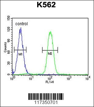
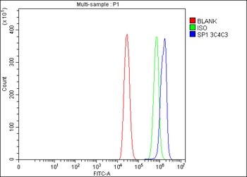
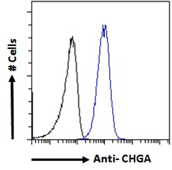
Flow cytometric analysis of K562 cells (right histogram) compared to a negative control cell (left histogram). FITC-conjugated goat-anti-rabbit secondary antibodies were used for the analysis.
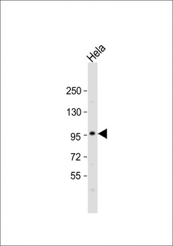
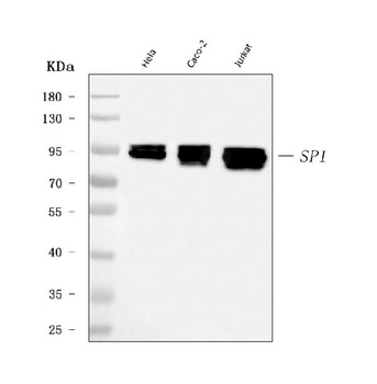
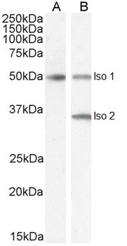
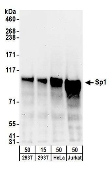
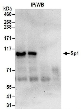
Western Blot at 1:500 dilution + Hela whole cell lysate Lysates/proteins at 20 ug per lane.
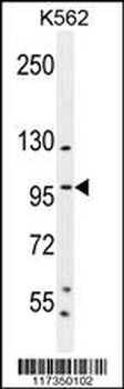
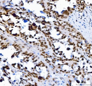
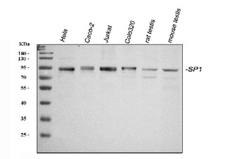
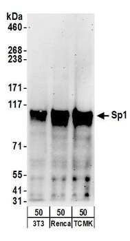
Western blot analysis in K562 cell line lysates (35 ug/lane).
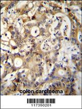
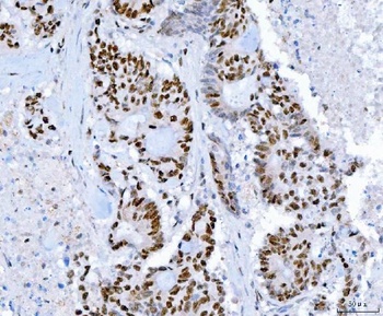
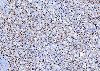
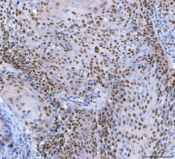
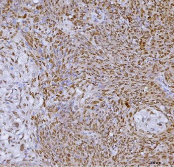
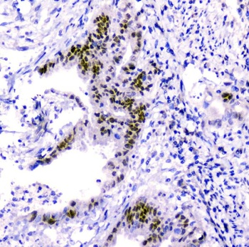
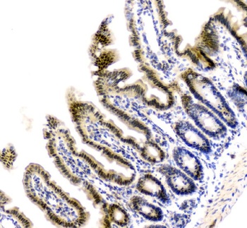
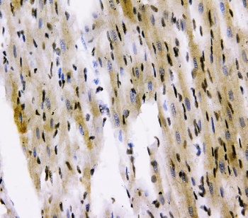
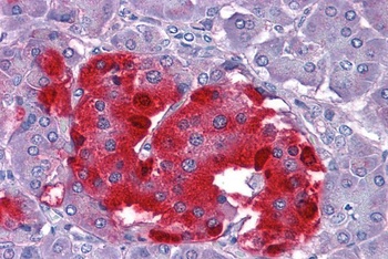
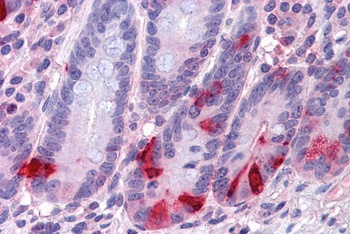
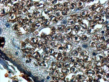
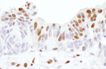
SP1 Antibody (C-term P692) immunohistochemistry analysis in formalin fixed and paraffin embedded human colon carcinoma followed by peroxidase conjugation of the secondary antibody and DAB staining.This data demonstrates the use o for immunohistochemistry.
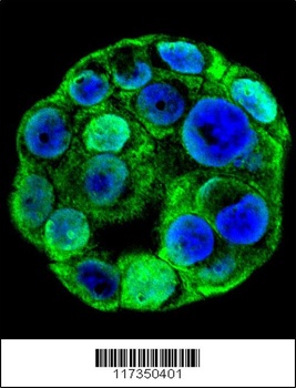
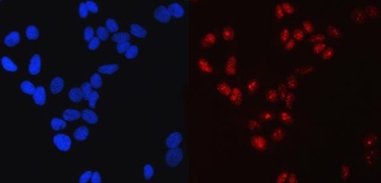
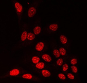
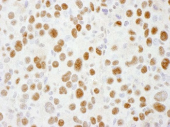
Confocal immunofluorescent analysis with WiDr cell followed by Alexa Fluor 488-conjugated goat anti-rabbit lgG (green). DAPI was used to stain the cell nuclear (blue).
- Item 1 of 10
SP1 Antibody (monoclonal, 3C4C3) [orb1474870]
FC, ICC, IF, IHC, WB
Human
Mouse
Monoclonal
Unconjugated
10 μg, 100 μg - Item 1 of 6
SP1 Antibody [orb443221]
ELISA, ICC, IF, IHC, WB
Human, Mouse, Rat
Rabbit
Polyclonal
Unconjugated
10 μg, 100 μg - Item 1 of 5
- Item 1 of 5
- Item 1 of 5


Submit a review
Filter by Rating

- 5 stars

- 4 stars
- 3 stars
- 2 stars
- 1 stars