You have no items in your shopping cart.
Featured
KO/KD
Validated
Validated
Description
Research Area
Infectious Disease & Virology
Images & Validation
−Item 1 of 9
| Tested Applications | ELISA, IF, IHC, KO/KD Validated, WB |
|---|---|
| Reactivity | Human, Mouse |
Key Properties
−| Antibody Type | Primary Antibody |
|---|---|
| Host | Rabbit |
| Clonality | Polyclonal |
| Isotype | IgG |
| Immunogen | Anti-SAMHD1 antibody (orb1239948) was raised against a peptide corresponding to 18 amino acids near the carboxy terminus of human SAMHD1. |
| Target | SAMHD1 |
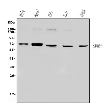
| Molecular Weight | Predicted: 72kDObserved: 72 kD |
| Purification | SAMHD1 antibody is affinity chromatography purified via peptide column. |
| Conjugation | Unconjugated |
Storage & Handling
−| Storage | Maintain refrigerated at 2-8°C for up to 2 weeks. For long term storage store at -20°C in small aliquots to prevent freeze-thaw cycles. |
|---|---|
| Form/Appearance | Liquid |
| Buffer/Preservatives | SAMHD1 antibody is supplied in PBS containing 0.02% sodium azide. |
| Concentration | 1 mg/mL |
| Expiration Date | 12 months from date of receipt. |
| Disclaimer | For research use only |
Alternative Names
−SAM domain and HD domain 1, DCIP, CHBL2, HDDC1, MOP-5, SBBI88
Similar Products
−- Item 1 of 14
SAMHD1 Mouse Monoclonal Antibody [orb738479]
FC, ICC, IF, IHC, WB
Human
Mouse
Monoclonal
Unconjugated
100 μg - Item 1 of 11
SAMHD1 Mouse Monoclonal Antibody [orb738478]
FC, ICC, IF, IHC, WB
Human
Mouse
Monoclonal
Unconjugated
100 μg - Item 1 of 12
SAMHD1 (phospho Thr592) Antibody [orb1239966]
ELISA, IF, IHC, KO/KD Validated, WB
Human, Mouse
Rabbit
Polyclonal
Unconjugated
0.1 mg, 0.02 mg - Item 1 of 7
SAMHD1 Rabbit Polyclonal Antibody [orb669122]
ELISA, FC, ICC, IF, IHC, WB
Human, Mouse, Rat
Rabbit
Polyclonal
Unconjugated
100 μg - Item 1 of 4
SAMHD1 rabbit pAb Antibody [orb767096]
ELISA, IF, IHC, WB
Human, Mouse, Rat
Polyclonal
Unconjugated
100 μl


Quality Guarantee
Explore bioreagents carefree to elevate your research. All our products are rigorously tested for performance. If a product does not perform as described on its datasheet, our scientific support team will provide expert troubleshooting, a prompt replacement, or a refund. For full details, please see our Terms & Conditions and Buying Guide. Contact us at [email protected].
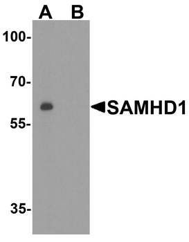
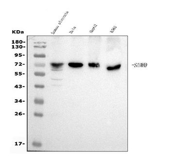
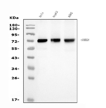
Western Blot Validation in Human Daudi Cell Lines. Loading: 15 µg of lysates per lane. Antibodies: SAMHD1 orb1239948, 1 µg/mL, in (A: the absence and B: the presence of blocking peptide), 1h incubation at RT in 5% NFDM/TBST. Secondary: Goat anti-rabbit IgG HRP conjugate at 1:10000 dilution.

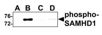
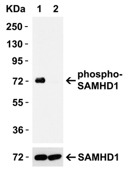
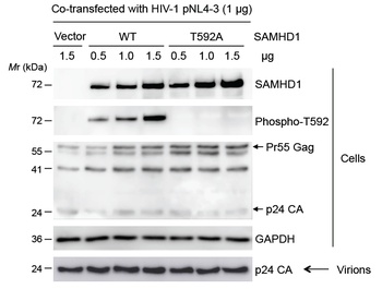
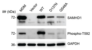
Overexpression Validation in 293T Transfected Cells. Loading: 15 µg of lysates per lane. Antibodies: SAMHD1 orb1239948 (0.1 µg/mL), (1h incubation at RT in 5% NFDM/TBST. Secondary: Goat anti-rabbit IgG HRP conjugate at 1:10000 dilution. 293 cells were transfected with (1) wild-type SAMHD1 or (2) SAMHD1 (mutation T592A).
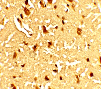
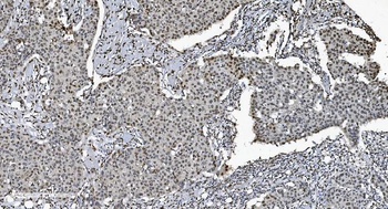
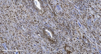
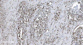
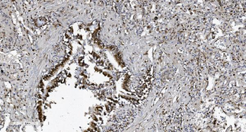
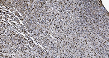
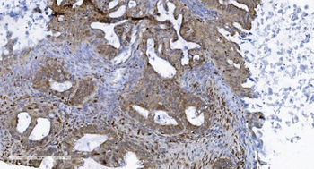
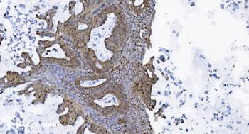
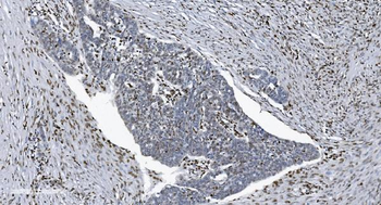
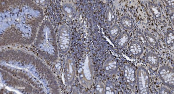
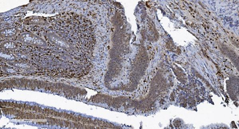
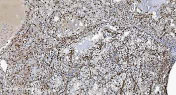
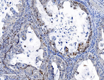
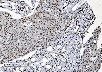
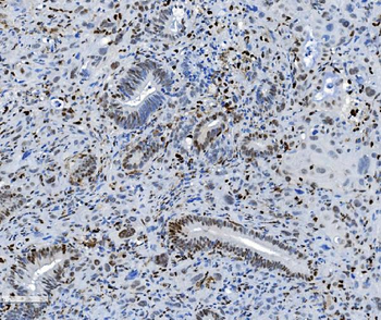
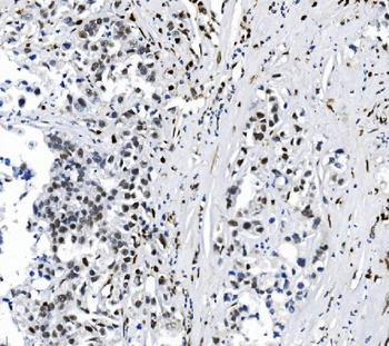
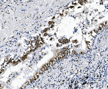
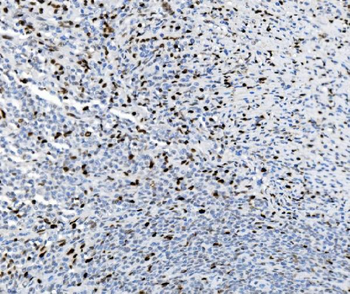
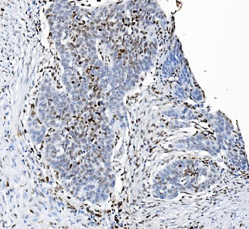
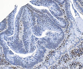
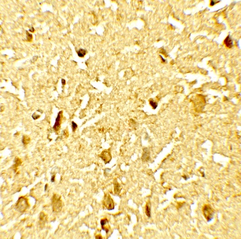
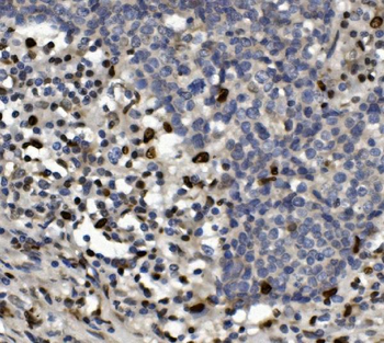
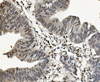
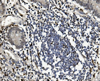
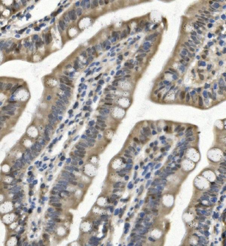
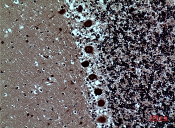
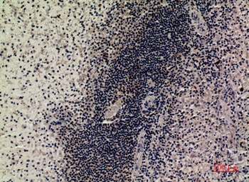
Immunohistochemistry Validation of SAMHD1 in Human Brain Tissue. Immunohistochemical analysis of paraffin-embedded Human Brain Tissue using anti-SAMHD1 antibody (orb1239948) at 5 µg/ml. Tissue was fixed with formaldehyde and blocked with 10% serum for 1 h at RT; antigen retrieval was by heat mediation with a citrate buffer (pH6). Samples were incubated with primary antibody overnight at 4 °C. A goat anti-rabbit IgG H&L (HRP) at 1/250 was used as secondary. Counter stained with Hematoxylin.
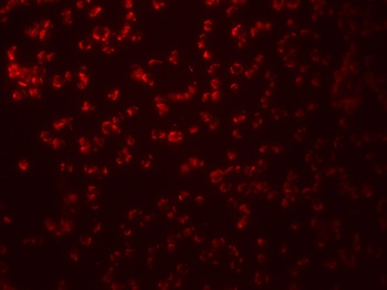
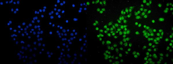
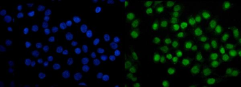
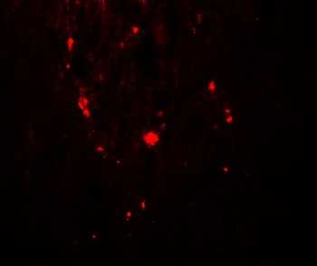
Immunofluorescence Validation of SAMHD1 in Human Daudi cells. Immunofluorescent analysis of 4% paraformaldehyde-fixed Human Daudi Cells labeling SAMHD1 with orb1239948 at 20 µg/mL, followed by goat anti-rabbit IgG secondary antibody at 1/500 dilution (red).
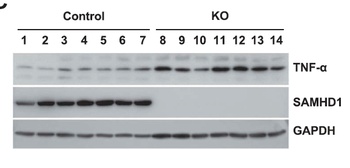
KO Validation of SAMHD1 in xenograft mice (Kodigepalli et al., 2018). THP-1 control and SAMHD1 KO (THP-1/KO) cells were injected into NSG (non-obese diabetic/severe combinedimmune deficient-gamma) mice. Protein expression levels of SAMHD1 were examined by Western blot with anti-SAMHD1 antibodies (orb1239948) and SAMHD1 was not detected in THP-1/KO cells.
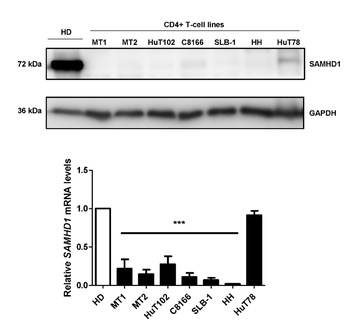
Overexpression Validation of SAMHD1 in CD4+ T-cells from a healthy donor and transformed CD4+ T-cell lines (Kohnken et al., 2017). MT1, MT2, SLB-1, and C8166 were from leukemia patients and HH, HuT78, and HuT102 were from cutaneous T-cell lymphoma (CTCL) patients. SAMHD1 protein expression detected by anti-SAMHD1 antibodies (orb1239948) was significantly increased in normal CD4+ T-cells as compared to leukemia- and CTCL- derived CD4+ T-cell lines. This blot is representative from four independent experiments with four healthy donors.
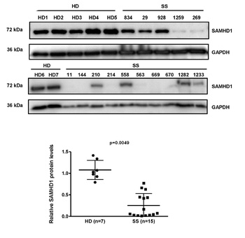
Overexpression of SAMHD1 in CD4+ T-cells from Healthy Donors and Sézary Syndrome (SS) patients (Kohnken et al., 2017). SAMHD1 protein expression detected by anti-SAMHD1 antibodies (orb1239948) was significantly reduced in CD+ T-cells from 15 SS patients as compared to those from 7 healthy donors.
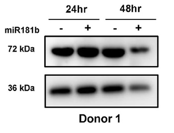
Regulated Expression Validation of SAMHD1 in CD4+ T-cells from a healthy donor (Kohnken et al., 2017). SAMHD1 protein expression detected by anti-SAMHD1 antibodies (orb1239948) was significantly decreased by about 40% relative to control cells at 48hr post-nucleofection with miR-181b.
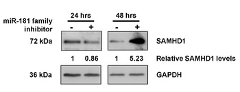
Regulated Expression Validation of SAMHD1 in MT2 CD4+ T-cells from leukemia patients (Kohnken et al., 2017). SAMHD1 protein expression detected by anti-SAMHD1 antibodies (orb1239948) was significantly increased by 5-fold at 48hr post-nucleofection with miR-181family inhibitor treatment.
Documents Download
Datasheet
Product Information
Request a Document
Protocol Information
WB
Western Blot (IB, immunoblot)
IHC
Immunohistochemistry
IF
Immunofluorescence
ELISA
Enzyme-linked Immunosorbent Assay (EIA)
SAMHD1 Antibody (orb1239948)

- 0.0
Based on 0 reviews
Participating in our Biorbyt product reviews program enables you to support fellow scientists by sharing your firsthand experience with our products.
Login to Submit a ReviewAvailable Sizes
Select a size below
Free Secondary Antibody (20 ul)0/0
Please add an antibody product to your cart first.