You have no items in your shopping cart.
Cart summary
Item 1 of 7
Item 1 of 7
PRKAA1 antibody
Catalog Number: orb49132

| Catalog Number | orb49132 |
|---|---|
| Category | Antibodies |
| Description | Rabbit polyclonal antibody to PRKAA1 |
| Species/Host | Rabbit |
| Clonality | Polyclonal |
| Tested applications | ELISA, IF, WB |
| Reactivity | Human, Mouse, Rat |
| Isotype | IgG |
| Immunogen | A synthetic peptide corresponding to a sequence within amino acids 381-520 of human AMPKα1 (NP_006242.5). |
| Concentration | 1 mg/ml |
| Dilution range | WB: 1:500-2000, IF/ICC: 1:50-200 |
| Purity | Affinity purification |
| Conjugation | Unconjugated |
| MW | 64kDa |
| Entrez | 5562 |
| UniProt ID | Q13131 |
| Storage | Maintain refrigerated at 2-8°C for up to 2 weeks. For long term storage store at -20°C in small aliquots to prevent freeze-thaw cycles. |
| Buffer/Preservatives | PBS with 0.01% thimerosal,50% glycerol,pH7.3. |
| Alternative names | anti PRKAA1 antibody, anti AMPK antibody, anti AMP Read more... |
| Note | For research use only |
| Expiration Date | 12 months from date of receipt. |

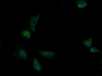
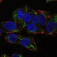
Immunofluorescence analysis of HeLa cells using AMPKα1 Rabbit pAb(orb49132) at a dilution of 1:100 (40x lens). Secondary antibody:Cy3 Goat Anti-Rabbit IgG (H+L) at 1:500 dilution. Blue: DAPI for nuclear staining.

Immunofluorescence analysis of MCF7 cells using AMPKα1 Rabbit pAb(orb49132) at a dilution of 1:100 (40x lens). Secondary antibody:Cy3 Goat Anti-Rabbit IgG (H+L) at 1:500 dilution. Blue: DAPI for nuclear staining.

Immunofluorescence analysis of PC-12 cells using AMPKα1 Rabbit pAb(orb49132) at a dilution of 1:100 (40x lens). Secondary antibody:Cy3 Goat Anti-Rabbit IgG (H+L) at 1:500 dilution. Blue: DAPI for nuclear staining.

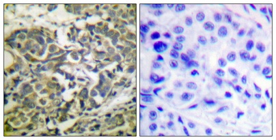
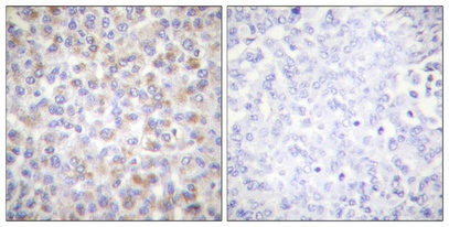
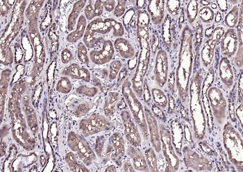
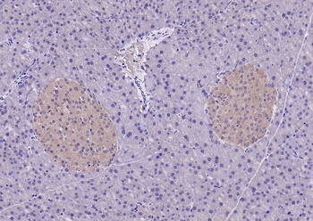
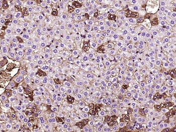
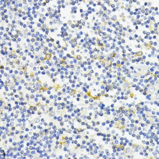
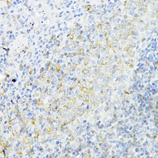
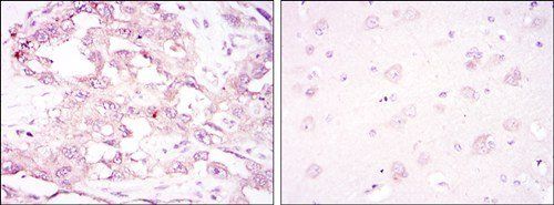
Immunohistochemistry analysis of paraffin-embedded Human liver tissue using AMPKα1 Rabbit pAb (orb49132) at a dilution of 1:300 (40x lens). High pressure antigen retrieval performed with 0.01M Citrate Bufferr (pH 6.0) prior to IHC staining.

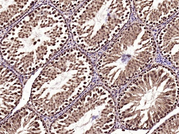
Immunohistochemistry analysis of paraffin-embedded Mouse testis tissue using AMPKα1 Rabbit pAb (orb49132) at a dilution of 1:300 (40x lens). High pressure antigen retrieval performed with 0.01M Citrate Bufferr (pH 6.0) prior to IHC staining.

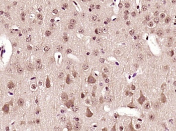
Immunohistochemistry analysis of paraffin-embedded Rat testis tissue using AMPKα1 Rabbit pAb (orb49132) at a dilution of 1:300 (40x lens). High pressure antigen retrieval performed with 0.01M Citrate Bufferr (pH 6.0) prior to IHC staining.

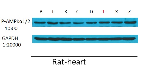
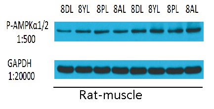
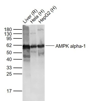
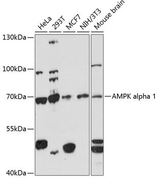
Western blot analysis of lysates from Rat liver, using AMPKα1 Rabbit pAb (orb49132) at 1:2000 dilution. Secondary antibody: HRP-conjugated Goat anti-Rabbit IgG (H+L) at 1:10000 dilution. Lysates/proteins: 25 µg per lane. Blocking buffer: 3% nonfat dry milk in TBST. Detection: ECL Basic Kit. Exposure time: 30s.
- Item 1 of 6
AMPK alpha 1(phospho-Thr172) antibody [orb99303]
ELISA, FC, IF, IHC-Fr, IHC-P, WB
Bovine, Canine, Equine, Gallus, Mouse, Porcine, Rat, Sheep
Human
Rabbit
Polyclonal
Unconjugated
50 μl, 100 μl, 200 μl - Item 1 of 6
AAPK1/AAPK2 Antibody [orb1563453]
ELISA, ICC, IHC-Fr, IHC-P, WB
Human, Monkey, Mouse, Porcine, Rat
Rabbit
Polyclonal
Unconjugated
100 μl, 50 μl, 20 μl - Item 1 of 6
AMPK Alpha-1 antibody [orb499641]
ELISA, IF, IHC-Fr, IHC-P, WB
Mouse, Rat
Human
Mouse
Monoclonal
Unconjugated
200 μl, 50 μl, 100 μl - Item 1 of 3
PRKAA1 antibody [orb541126]
ELISA, IHC-P, WB
Human, Mouse, Rat
Rabbit
Polyclonal
Unconjugated
50 μl, 100 μl, 200 μl - Item 1 of 5
PRKAA1 Antibody [orb69386]
ELISA, FC, ICC, IHC, WB
Human, Monkey, Mouse, Rat
Mouse
Monoclonal
Unconjugated
100 μl
Submit a review
Filter by Rating

- 5 stars

- 4 stars
- 3 stars
- 2 stars
- 1 stars
