You have no items in your shopping cart.
Cart summary
Item 1 of 5
Item 1 of 5
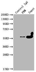
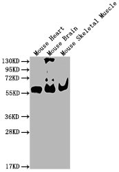
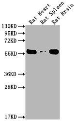
PKM Antibody
Catalog Number: orb1271022
| Catalog Number | orb1271022 |
|---|---|
| Category | Antibodies |
| Description | PKM Antibody |
| Species/Host | Rabbit |
| Clonality | Polyclonal |
| Tested applications | IF, IHC-P, WB |
| Reactivity | Human, Monkey, Mouse, Rat |
| Isotype | Rabbit Ig |
| Immunogen | This Pyruvate Kinase (PKM2) antibody is generated from rabbits immunized with a KLH conjugated synthetic peptide between 476-505 amino acids from the C-terminal region of human Pyruvate Kinase (PKM2). |
| Concentration | batch dependent |
| Dilution range | For WB starting dilution is: 1:1000For IF starting dilution is: 1:200For IHC-P starting dilution is: 1:50~1:100 |
| Form/Appearance | Liquid |
| Conjugation | Unconjugated |
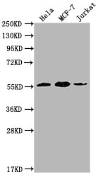
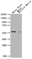
| MW | 58 kDa |
| Target | PKM |
| UniProt ID | P14618 |
| NCBI | P14618 |
| Storage | Store at 4°C for three months and -20°C, stable for up to one year. As with all antibodies care should be taken to avoid repeated freeze thaw cycles. Antibodies should not be exposed to prolonged high temperatures. |
| Buffer/Preservatives | Supplied in PBS with 0.09% (W/V) sodium azide. |
| Alternative names | Pyruvate kinase PKM, Cytosolic thyroid hormone-bin Read more... |
| Note | For research use only |
| Application notes | For WB starting dilution is: 1:1000For IF starting dilution is: 1:200For IHC-P starting dilution is: 1:50~1:100 |
| Expiration Date | 12 months from date of receipt. |
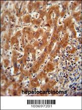
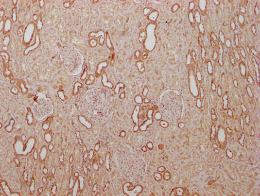
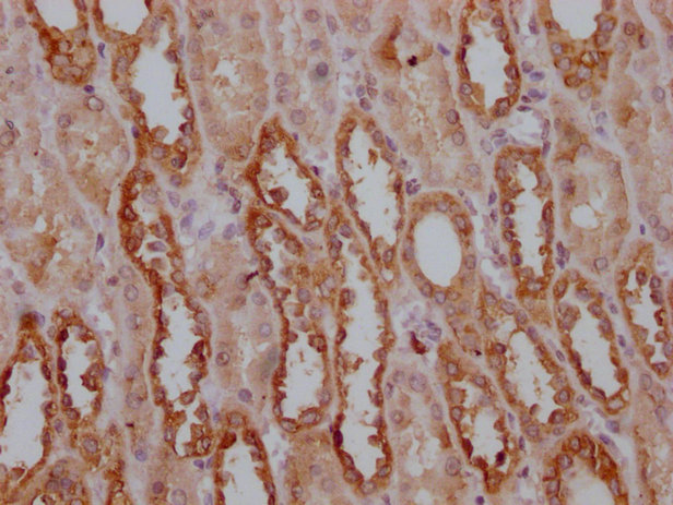
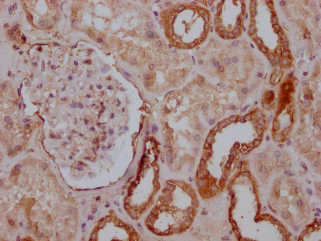
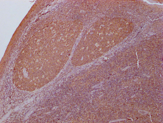
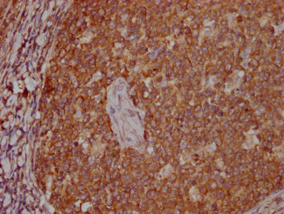
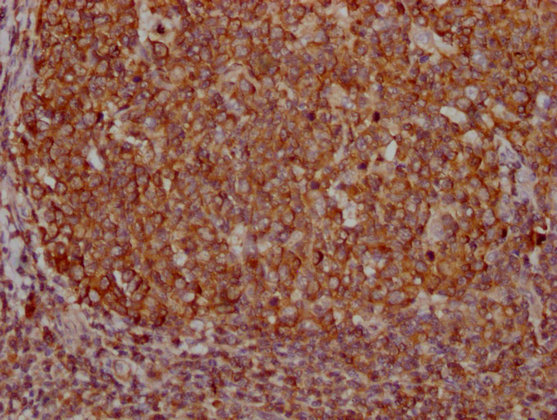
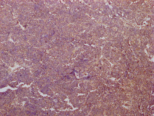
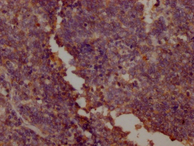
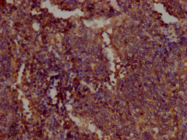
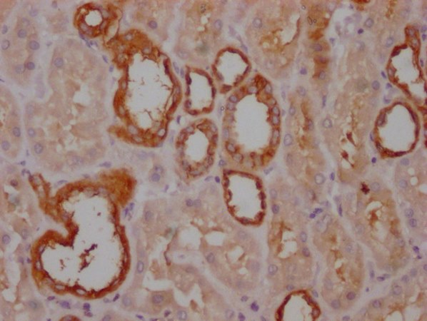
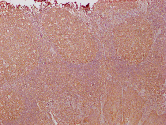
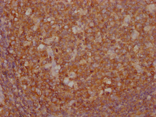
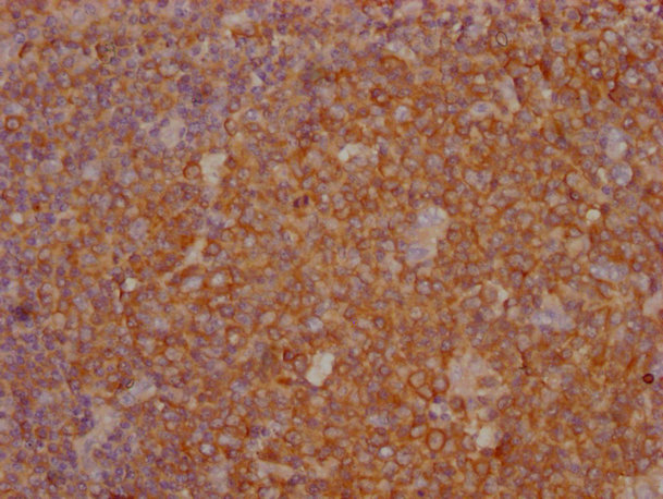
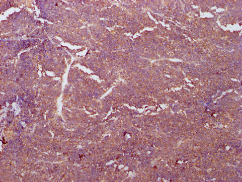
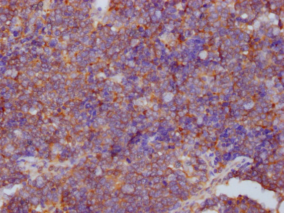
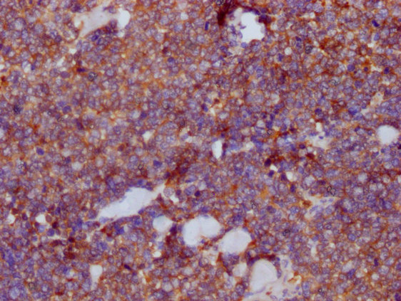
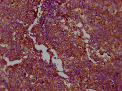
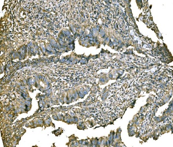
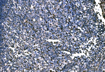
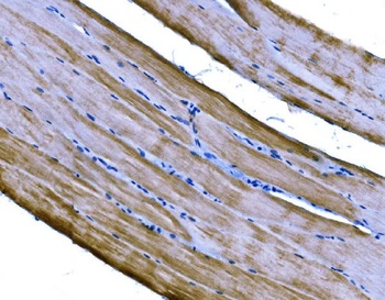
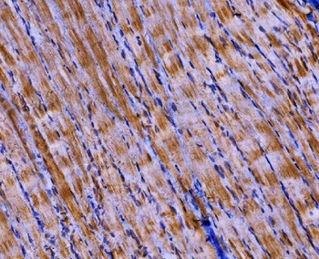
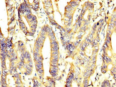
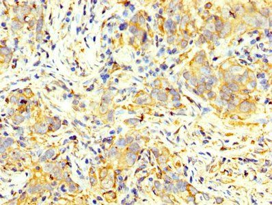
Pyruvate Kinase (PKM2) Antibody immunohistochemistry analysis in formalin fixed and paraffin embedded human hepatocarcinoma followed by peroxidase conjugation of the secondary antibody and DAB staining. This data demonstrates the use of the Pyruvate Kinase (PKM2) Antibody for immunohistochemistry.
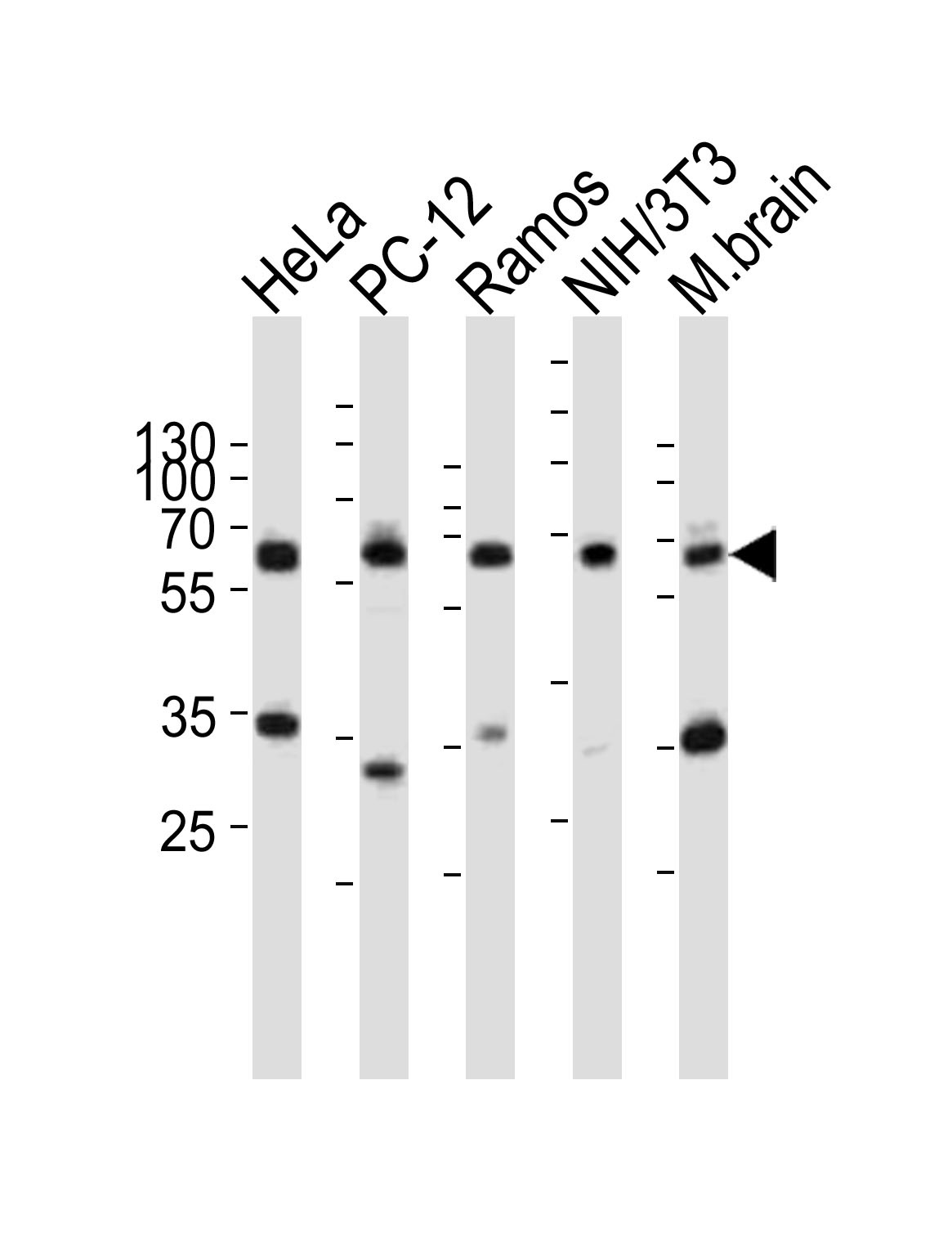
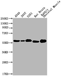
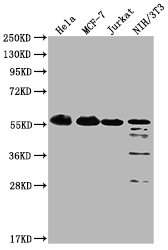
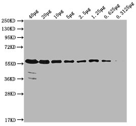
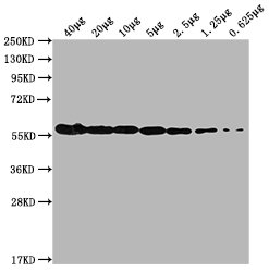
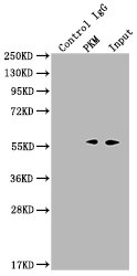
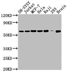
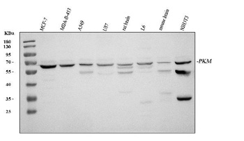
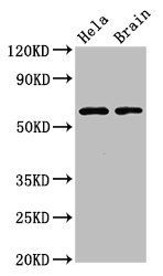
Western blot analysis of lysates from HeLa, rat PC-12, Ramos, mouse NIH/3T3 cell line, mouse brain tissue lysate (from left to right), using PKM2-N491 at 1:1000 at each lane.
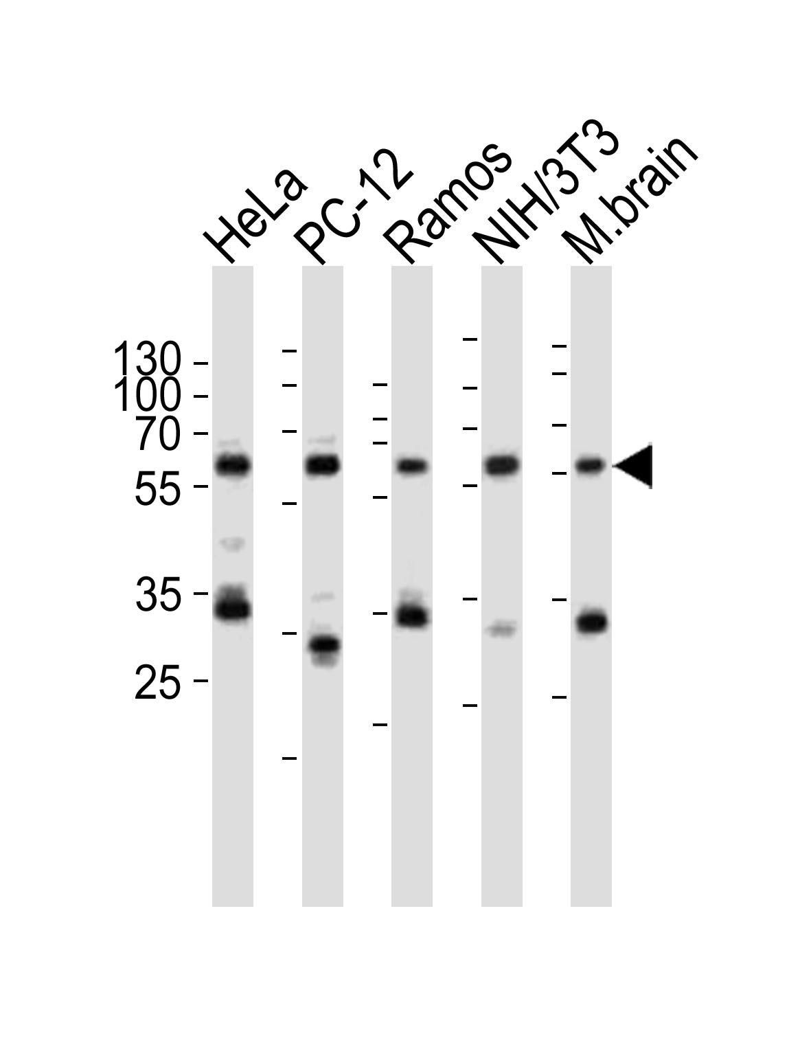
Western blot analysis of lysates from HeLa, rat PC-12, Ramos, mouse NIH/3T3 cell line, mouse brain tissue lysate (from left to right), using PKM2-N491 at 1:1000 at each lane.
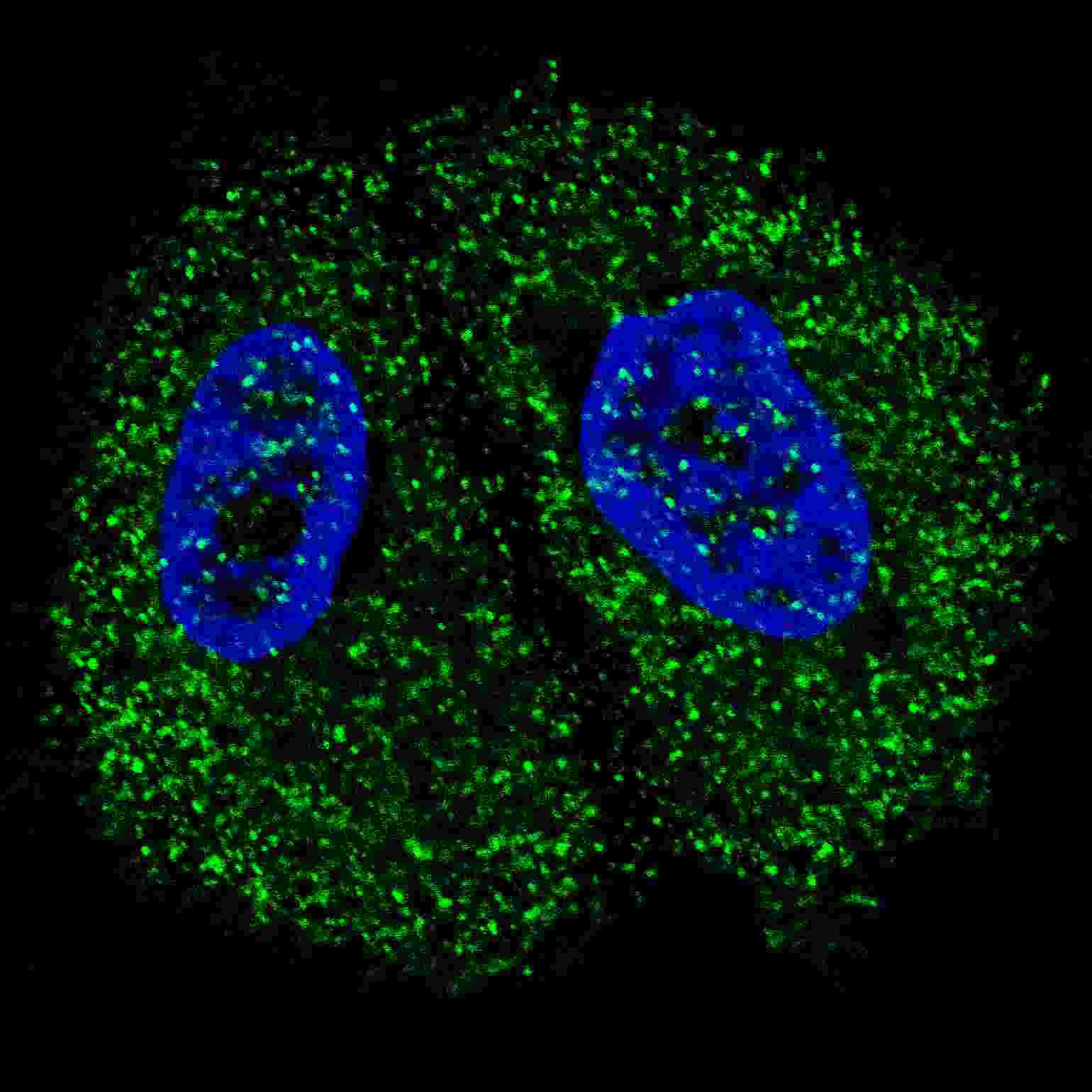
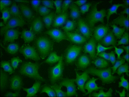
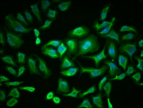
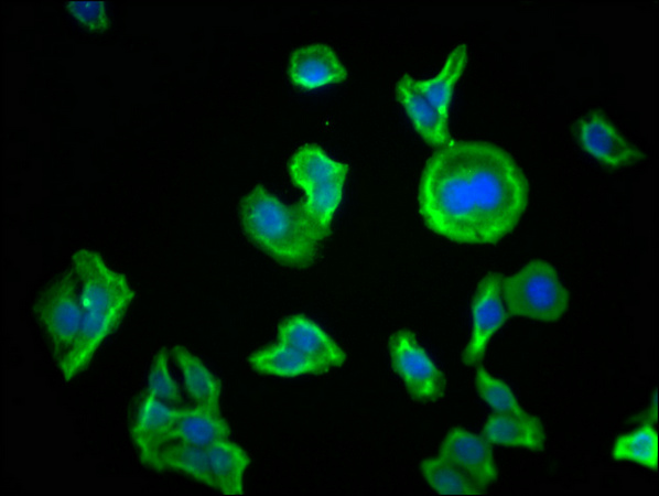
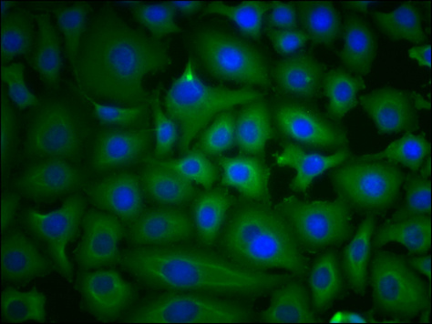
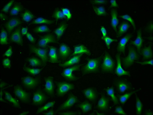
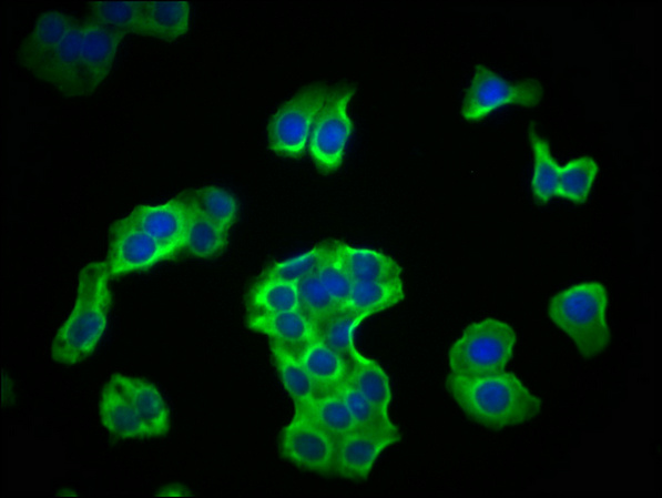
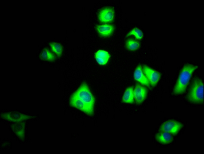
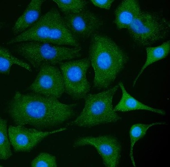
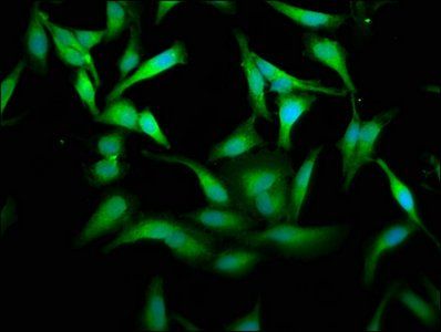
Fluorescent confocal image of MCF7 cells stained with Pyruvate Kinase (PKM2) antibody. MCF7 cells were fixed with 4% PFA (20 min), permeabilized with Triton X-100 (0.2%, 30 min). Cells were then incubated with Pyruvate Kinase (PKM2) primary antibody (1:200, 2 h at room temperature). For secondary antibody, Alexa Fluor 488 conjugated donkey anti-rabbit antibody (green) was used (1:1000, 1h). Nuclei were counterstained with Hoechst 33342 (blue) (10 ug/ml, 5 min). Note the highly specific localization of the Pyruvate Kinase (PKM2) mainly to the cytoplasm, supported by Human Protein Atlas Data (http://www.proteinatlas.org/ENSG00000067225).
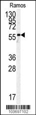
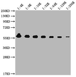
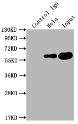
Western blot analysis of anti-h PKM2-N491 Pab in Ramos cell line lysates (35 ug/lane).
- Item 1 of 21
PKM antibody [orb688872]
ELISA, FC, IF, IHC, IP, WB
Human, Mouse, Rabbit, Rat
Mouse
Monoclonal
Unconjugated
100 μl, 50 μl - Item 1 of 19
PKM antibody [orb688873]
ELISA, FC, IF, IHC, IP, WB
Human, Mouse
Mouse
Monoclonal
Unconjugated
100 μl, 50 μl - Item 1 of 5
PKM antibody [orb688982]
ELISA, FC, IF, IHC, IP, WB
Human, Mouse
Rabbit
Monoclonal
Unconjugated
100 μl, 50 μl - Item 1 of 6
PKM2 Antibody [orb251562]
ICC, IF, IHC, WB
Bovine
Human, Mouse, Rat
Rabbit
Polyclonal
Unconjugated
10 μg, 100 μg - Item 1 of 4

Submit a review
Filter by Rating

- 5 stars

- 4 stars
- 3 stars
- 2 stars
- 1 stars