You have no items in your shopping cart.
Page Not Found
Cart summary
Item 1 of 5
Item 1 of 5
PIM2 Antibody
Catalog Number: orb1262853

| Catalog Number | orb1262853 |
|---|---|
| Category | Antibodies |
| Description | PIM2 Antibody |
| Species/Host | Rabbit |
| Clonality | Polyclonal |
| Tested applications | IHC-P, IP, WB |
| Reactivity | Human |
| Isotype | Rabbit Ig |
| Immunogen | This PIM2 antibody is generated from rabbits immunized with a KLH conjugated synthetic peptide between 277-308 amino acids from the C-terminal region of human PIM2. |
| Antibody Type | Primary Antibody |
| Concentration | batch dependent |
| Form/Appearance | Liquid |
| Conjugation | Unconjugated |
| MW | 34 kDa |
| Target | PIM2 |
| UniProt ID | Q9P1W9 |
| NCBI | Q9P1W9 |
| Storage | Maintain refrigerated at 2-8°C for up to 2 weeks. For long term storage store at -20°C in small aliquots to prevent freeze-thaw cycles. |
| Buffer/Preservatives | Supplied in PBS with 0.09% (W/V) sodium azide. |
| Alternative names | Serine/threonine-protein kinase pim-2, Pim-2h, PIM Read more... |
| Note | For research use only |
| Application notes | For WB starting dilution is: 1:1000For IP starting dilution is: 1:50~100For IHC-P starting dilution is: 1:50~100 |
| Expiration Date | 12 months from date of receipt. |
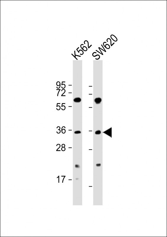
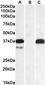
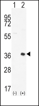
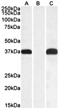
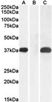
Western Blot at 1:2000 dilution Lane 1: K562 whole cell lysate Lane 2: SW620 whole cell lysate Lysates/proteins at 20 ug per lane.
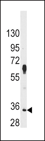
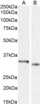
Western blot analysis of anti-PIM2 Antibody in Hela cell line lysates (35 ug/lane)

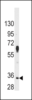
Western blot analysis of PIM2 using rabbit polyclonal PIM2 Antibody (D292) using 293 cell lysates (2 ug/lane) either nontransfected (Lane 1) or transiently transfected (Lane 2) with the PIM2 gene.

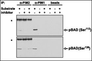
PIM proteins were immunoprecipitated from MV4;11 cells and the agarose-protein A-immunoprecipitate complex was tested for its ability to phosphorylate BAD in vitro in the presence or absence of K00135. Phosphorylation of BAD (both on Ser112 and Ser136, detected by WB with phospho-specific antibodies) was abrogated on addition of the compound. Asterisks, strong bands corresponding to the heavy chain of the anti-PIM2 rabbit antibody recognized by the antirabbit immunoglobulin G secondary antibody. Beads alone (without anti-PIM antibodies) were incubated with the MV4;11 extract and used for the same in vitro phosphorylation reaction as a negative control.
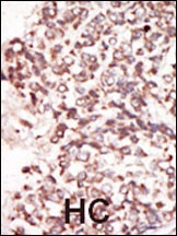
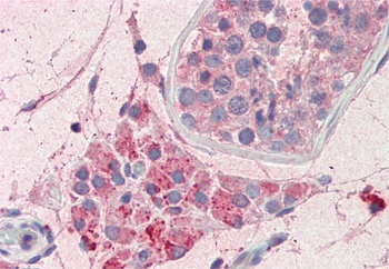
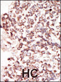
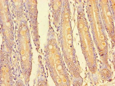
Formalin-fixed and paraffin-embedded human cancer tissue reacted with the primary antibody, which was peroxidase-conjugated to the secondary antibody, followed by DAB staining. BC = breast carcinoma; HC = hepatocarcinoma.
- Item 1 of 5
- Item 1 of 5
- Item 1 of 3
- Item 1 of 1
Goat anti-PIM2 (aa23-36) Antibody [orb131689]
ELISA, IHC, WB
Bovine, Human, Mouse, Rat
Goat
Polyclonal
Unconjugated
100 μg - Item 1 of 1
Goat anti-PIM2 (Internal) Antibody [orb137118]
ELISA, WB
Bovine, Canine, Human, Mouse
Goat
Polyclonal
Unconjugated
100 μg