You have no items in your shopping cart.
Cart summary
Item 1 of 2
Item 1 of 2
PHGDH antibody
Catalog Number: orb411781
| Catalog Number | orb411781 |
|---|---|
| Category | Antibodies |
| Description | Rabbit polyclonal antibody to PHGDH |
| Species/Host | Rabbit |
| Clonality | Polyclonal |
| Tested applications | IF, WB |
| Reactivity | Human, Mouse, Rat |
| Immunogen | Recombinant full length protein of human PGDH3 |
| Dilution range | WB: 1:500-2000 |
| Form/Appearance | Liquid in 0.42% Potassium phosphate, 0.87% Sodium chloride, pH 7.3, 30% glycerol, and 0.01% sodium azide. |
| Conjugation | Unconjugated |
| Target | PHGDH |
| Entrez | 236539, 26227, 58835 |
| UniProt ID | O43175, Q61753, O08651 |
| Source | Rabbit |
| Storage | Shipped at 4°C. Upon delivery aliquot and store at -20°C for one year. Avoid freeze/thaw cycles. |
| Buffer/Preservatives | Liquid in 0.42% Potassium phosphate, 0.87% Sodium chloride, pH 7.3, 30% glycerol, and 0.01% sodium azide. |
| Alternative names | anti-PGDH3 antibody, anti-D-3-phosphoglycerate deh Read more... |
| Note | For research use only |
| Expiration Date | 12 months from date of receipt. |
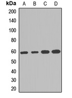
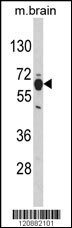
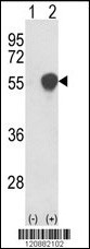
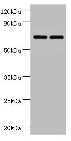
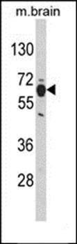
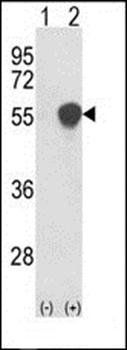
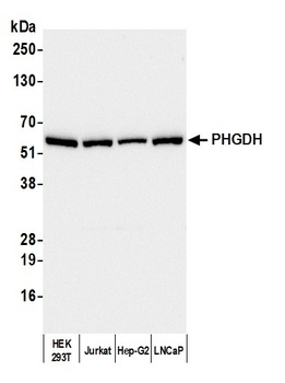
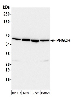
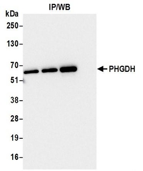
Western blot analysis of PGDH3 expression in Hela (A), SKOV3 (B), mouse brain (C), rat spinal cord (D) whole cell lysates. (Predicted band size: 56 kD; Observed band size: 59 kD)
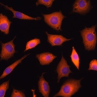
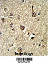
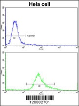
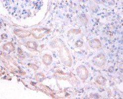
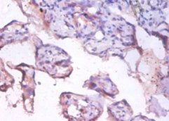
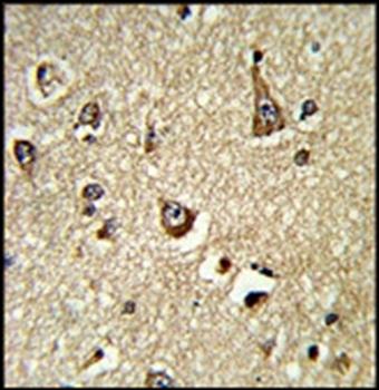
Immunofluorescent analysis of PGDH3 staining in L929 cells. Formalin-fixed cells were permeabilized with 0.1% Triton X-100 in TBS for 5-10 minutes and blocked with 3% BSA-PBS for 30 minutes at room temperature. Cells were probed with the primary antibody in 3% BSA-PBS and incubated overnight at 4 °C in a humidified chamber. Cells were washed with PBST and incubated with a AF594-conjugated secondary antibody (red) in PBS at room temperature in the dark. DAPI was used to stain the cell nuclei (blue).
- Item 1 of 5
- Item 1 of 4
PHGDH Antibody [orb1264496]
FC, IHC-P, WB
Bovine, Monkey, Porcine
Human, Mouse
Rabbit
Polyclonal
Unconjugated
400 μl - Item 1 of 3
D-3-phosphoglycerate dehydrogenase antibody [orb239893]
ELISA, IHC, WB
Human
Rabbit
Polyclonal
Unconjugated
100 μg, 50 μg - Item 1 of 4
- Item 1 of 4


Submit a review
Filter by Rating

- 5 stars

- 4 stars
- 3 stars
- 2 stars
- 1 stars