You have no items in your shopping cart.
Cart summary
Item 1 of 5
Item 1 of 5
PC4/SUB1 Antibody
Catalog Number: orb865652
| Catalog Number | orb865652 |
|---|---|
| Category | Antibodies |
| Description | PC4/SUB1 Antibody |
| Species/Host | Rabbit |
| Clonality | Polyclonal |
| Tested applications | ELISA, IF, IHC, WB |
| Reactivity | Human, Mouse, Rat |
| Isotype | Rabbit IgG |
| Immunogen | E.coli-derived human PC4/SUB1 recombinant protein (Position: N62-L127). |
| Concentration | Adding 0.2 ml of distilled water will yield a concentration of 500 μg/ml. |
| Dilution range | Western blot, 0.1-0.25 μg/ml, Human, Mouse, Rat Immunohistochemistry(Paraffin-embedded Section), 2-5 μg/ml, Human Immunofluorescence, 5 μg/ml, Human Direct ELISA, 0.1-0.5 μg/ml, Human |
| Form/Appearance | Lyophilized |
| Conjugation | Unconjugated |
| MW | 19 kDa |
| UniProt ID | P53999 |
| Storage | At -20°C for one year from date of receipt. After reconstitution, at 4°C for one month. It can also be aliquotted and stored frozen at -20°C for six months. Avoid repeated freezing and thawing. |
| Note | For research use only |
| Application notes | Tested Species: In-house tested species with positive results. Other applications have not been tested. Optimal dilutions should be determined by end users. Adding 0.2 ml of distilled water will yield a concentration of 500 μg/ml. |
| Expiration Date | 12 months from date of receipt. |
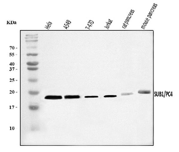
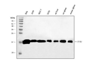
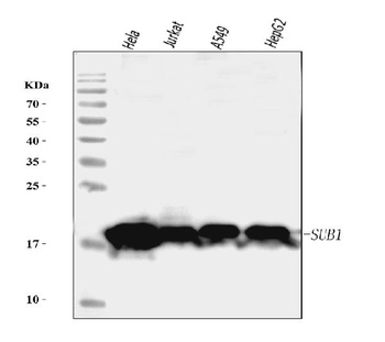
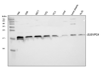
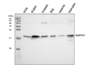
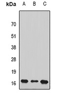
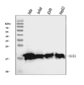
Western blot analysis of PC4/SUB1 using anti-PC4/SUB1 antibody (orb865652). Electrophoresis was performed on a 5-20% SDS-PAGE gel at 70V (Stacking gel)/90V (Resolving gel) for 2-3 hours. The sample well of each lane was loaded with 30 ug of sample under reducing conditions. Lane 1: human HeLa whole cell lysates, Lane 2: human A549 whole cell lysates, Lane 3: human T-47D whole cell lysates, Lane 4: human Jurkat whole cell lysates, Lane 5: rat pancreas tissue lysates, Lane 6: mouse pancreas tissue lysates. After electrophoresis, proteins were transferred to a nitrocellulose membrane at 150 mA for 50-90 minutes. Blocked the membrane with 5% non-fat milk/TBS for 1.5 hour at RT. The membrane was incubated with rabbit anti-PC4/SUB1 antigen affinity purified polyclonal antibody (Catalog # orb865652) at 0.25 μg/mL overnight at 4°C, then washed with TBS-0.1%Tween 3 times with 5 minutes each and probed with a goat anti-rabbit IgG-HRP secondary antibody at a dilution of 1:5000 for 1.5 hour at RT. The signal is developed using an Enhanced Chemiluminescent detection (ECL) kit (Catalog # orb90503) with Tanon 5200 system. A specific band was detected for PC4/SUB1 at approximately 19 kDa. The expected band size for PC4/SUB1 is at 14 kDa.
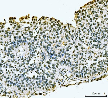
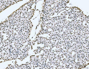
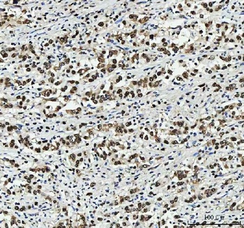
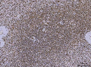
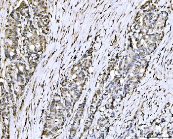
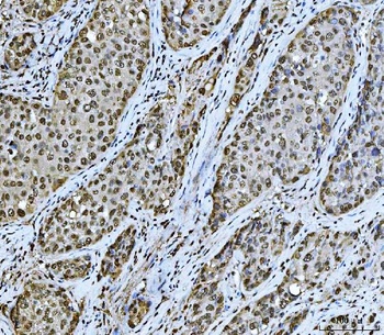
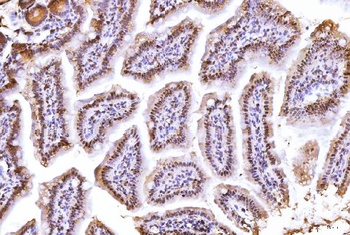
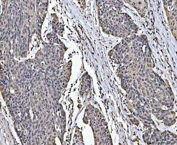
IHC analysis of PC4/SUB1 using anti-PC4/SUB1 antibody (orb865652). PC4/SUB1 was detected in a paraffin-embedded section of human bladder epithelial carcinoma tissue. Heat mediated antigen retrieval was performed in EDTA buffer (pH 8.0, epitope retrieval solution). The tissue section was blocked with 10% goat serum. The tissue section was then incubated with 2 μg/ml rabbit anti-PC4/SUB1 Antibody (orb865652) overnight at 4°C. Biotinylated goat anti-rabbit IgG was used as secondary antibody and incubated for 30 minutes at 37°C. The tissue section was developed using Strepavidin-Biotin-Complex (SABC) (Catalog # orb90444) with DAB as the chromogen.
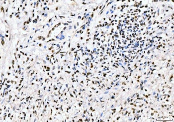
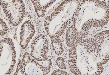
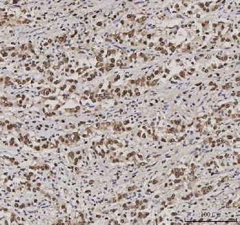
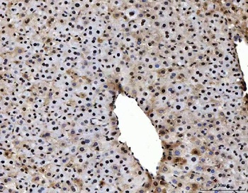
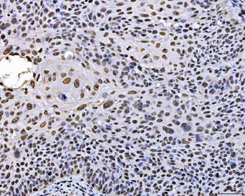
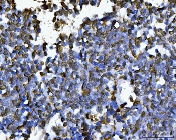
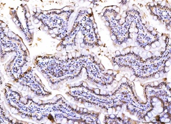
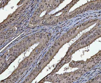
IHC analysis of PC4/SUB1 using anti-PC4/SUB1 antibody (orb865652). PC4/SUB1 was detected in a paraffin-embedded section of human gastric signet ring cell carcinoma tissue. Heat mediated antigen retrieval was performed in EDTA buffer (pH 8.0, epitope retrieval solution). The tissue section was blocked with 10% goat serum. The tissue section was then incubated with 2 μg/ml rabbit anti-PC4/SUB1 Antibody (orb865652) overnight at 4°C. Biotinylated goat anti-rabbit IgG was used as secondary antibody and incubated for 30 minutes at 37°C. The tissue section was developed using Strepavidin-Biotin-Complex (SABC) (Catalog # orb90444) with DAB as the chromogen.
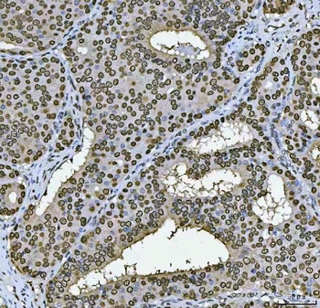
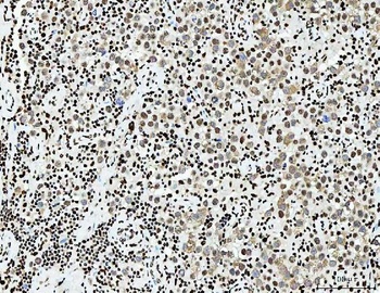
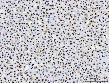
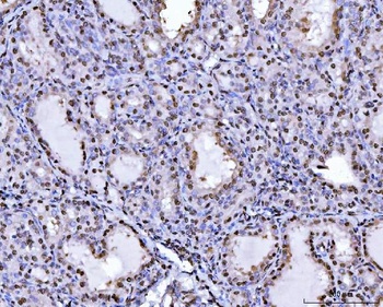
IHC analysis of PC4/SUB1 using anti-PC4/SUB1 antibody (orb865652). PC4/SUB1 was detected in a paraffin-embedded section of human gastric signet ring cell carcinoma tissue. Heat mediated antigen retrieval was performed in EDTA buffer (pH 8.0, epitope retrieval solution). The tissue section was blocked with 10% goat serum. The tissue section was then incubated with 2 μg/ml rabbit anti-PC4/SUB1 Antibody (orb865652) overnight at 4°C. Biotinylated goat anti-rabbit IgG was used as secondary antibody and incubated for 30 minutes at 37°C. The tissue section was developed using Strepavidin-Biotin-Complex (SABC) (Catalog # orb90444) with DAB as the chromogen.
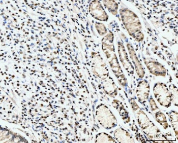
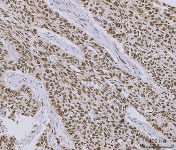
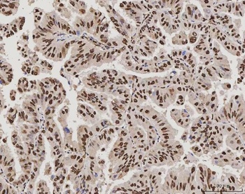
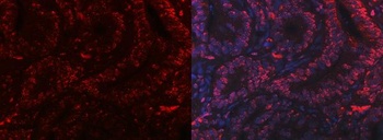
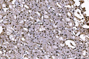
IHC analysis of PC4/SUB1 using anti-PC4/SUB1 antibody (orb865652). PC4/SUB1 was detected in a paraffin-embedded section of human papillary carcinoma of the left breast tissue. Heat mediated antigen retrieval was performed in EDTA buffer (pH 8.0, epitope retrieval solution). The tissue section was blocked with 10% goat serum. The tissue section was then incubated with 2 μg/ml rabbit anti-PC4/SUB1 Antibody (orb865652) overnight at 4°C. Biotinylated goat anti-rabbit IgG was used as secondary antibody and incubated for 30 minutes at 37°C. The tissue section was developed using Strepavidin-Biotin-Complex (SABC) (Catalog # orb90444) with DAB as the chromogen.
- Item 1 of 13
PC4/SUB1 Antibody (monoclonal, 8D9D1) [orb1184747]
FC, IF, IHC, WB
Human, Mouse, Rat
Mouse
Monoclonal
Unconjugated
10 μg, 100 μg - Item 1 of 9
PC4/SUB1 Antibody (monoclonal, 6B5B10) [orb1145777]
IHC, WB
Human, Mouse, Rat
Mouse
Monoclonal
Unconjugated
10 μg, 100 μg - Item 1 of 5
PC4/SUB1 Antibody [orb381100]
IF, IHC, WB
Hamster
Human, Mouse, Rat
Rabbit
Polyclonal
Unconjugated
10 μg, 100 μg - Item 1 of 2
- Item 1 of 2
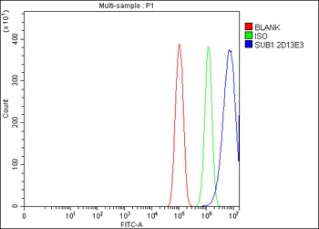
PC4/SUB1 Antibody (monoclonal, 2D13E3) [orb1145778]
FC, WB
Human
Mouse
Monoclonal
Unconjugated
10 μg, 100 μg
Submit a review
Filter by Rating

- 5 stars

- 4 stars
- 3 stars
- 2 stars
- 1 stars