You have no items in your shopping cart.
Cart summary
Item 1 of 6
Item 1 of 6
PACSIN2 Antibody
Catalog Number: orb1262761

| Catalog Number | orb1262761 |
|---|---|
| Category | Antibodies |
| Description | PACSIN2 Antibody |
| Target | PACSIN2 |
| Clonality | Polyclonal |
| Isotype | Rabbit Ig |
| Conjugation | Unconjugated |
| Reactivity | Human, Mouse |
| Form/Appearance | Liquid |
| Concentration | batch dependent |
| Buffer/Preservatives | Supplied in PBS with 0.09% (W/V) sodium azide. |
| Immunogen | This PACSIN2 antibody is generated from rabbits immunized with a KLH conjugated synthetic peptide between 342-371 amino acids from the C-terminal region of human PACSIN2. |
| UniProt ID | Q9UNF0 |
| MW | 56 kDa |
| Tested applications | IF, IHC-P, WB |
| Application notes | For IHC-P starting dilution is: 1:25For IF starting dilution is: 1:100For WB starting dilution is: 1:1000 |
| Antibody Type | Primary Antibody |
| Storage | Maintain refrigerated at 2-8°C for up to 2 weeks. For long term storage store at -20°C in small aliquots to prevent freeze-thaw cycles. |
| Alternative names | Protein kinase C and casein kinase substrate in ne Read more... |
| Note | For research use only |
| NCBI | Q9UNF0 |
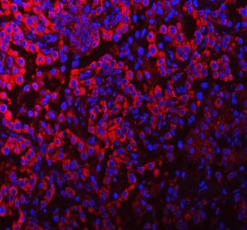
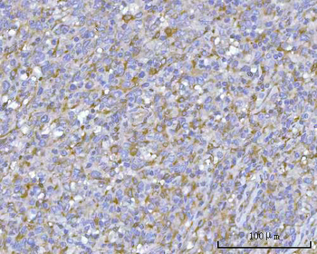
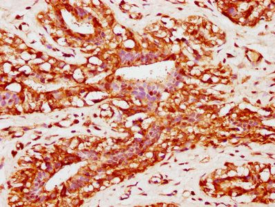
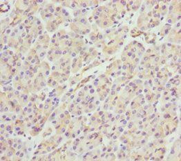
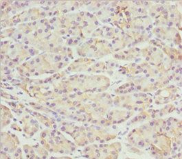
Immunohistochemical analysis of paraffin-embedded H. stomach section using PACSIN2 Antibody. Antibody was diluted at 1:25 dilution. A peroxidase-conjugated goat anti-rabbit IgG at 1:400 dilution was used as the secondary antibody, followed by DAB staining.
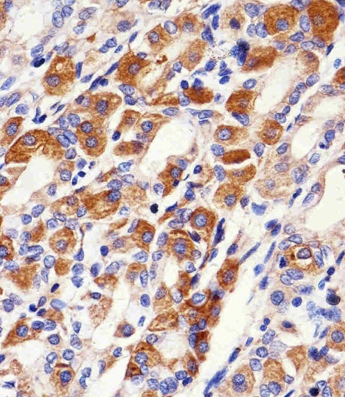
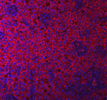
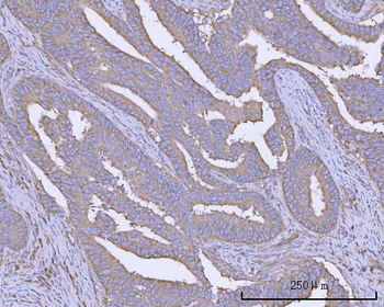
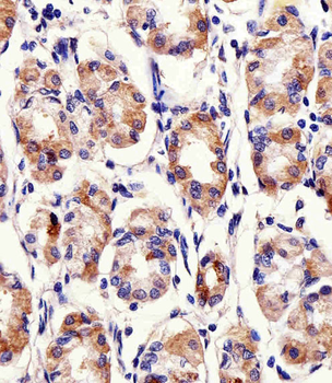
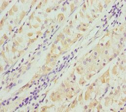
Immunohistochemical analysis of paraffin-embedded H.stomach section using PACSIN2 Antibody. Antibody was diluted at 1:100 dilution. A peroxidase-conjugated goat anti-rabbit IgG at 1:400 dilution was used as the secondary antibody, followed by DAB staining.
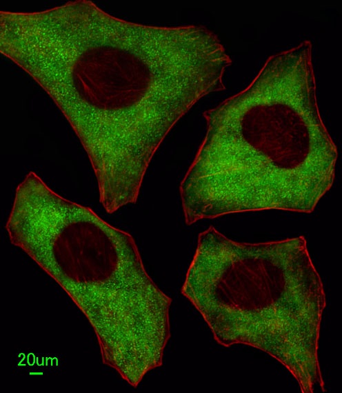
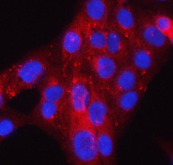
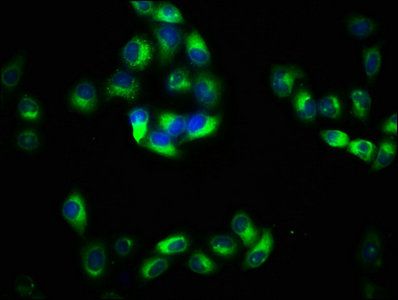
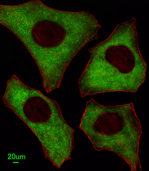
Immunofluorescent analysis of Hela cells, using PACSIN2 Antibody. Antibody was diluted at 1:100 dilution. Alexa Fluor 488-conjugated goat anti-rabbit lgG at 1:400 dilution was used as the secondary antibody (green).Cytoplasmic actin was counterstained with Fluor 554 (red) conjugated Phalloidin (red).

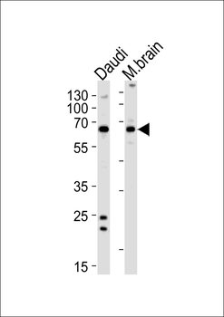
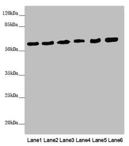
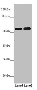
Western blot analysis in Daudi cell line and mouse brain lysates (35 ug/lane).This demonstrates the PACSIN2 antibody detected the PACSIN2 protein (arrow).
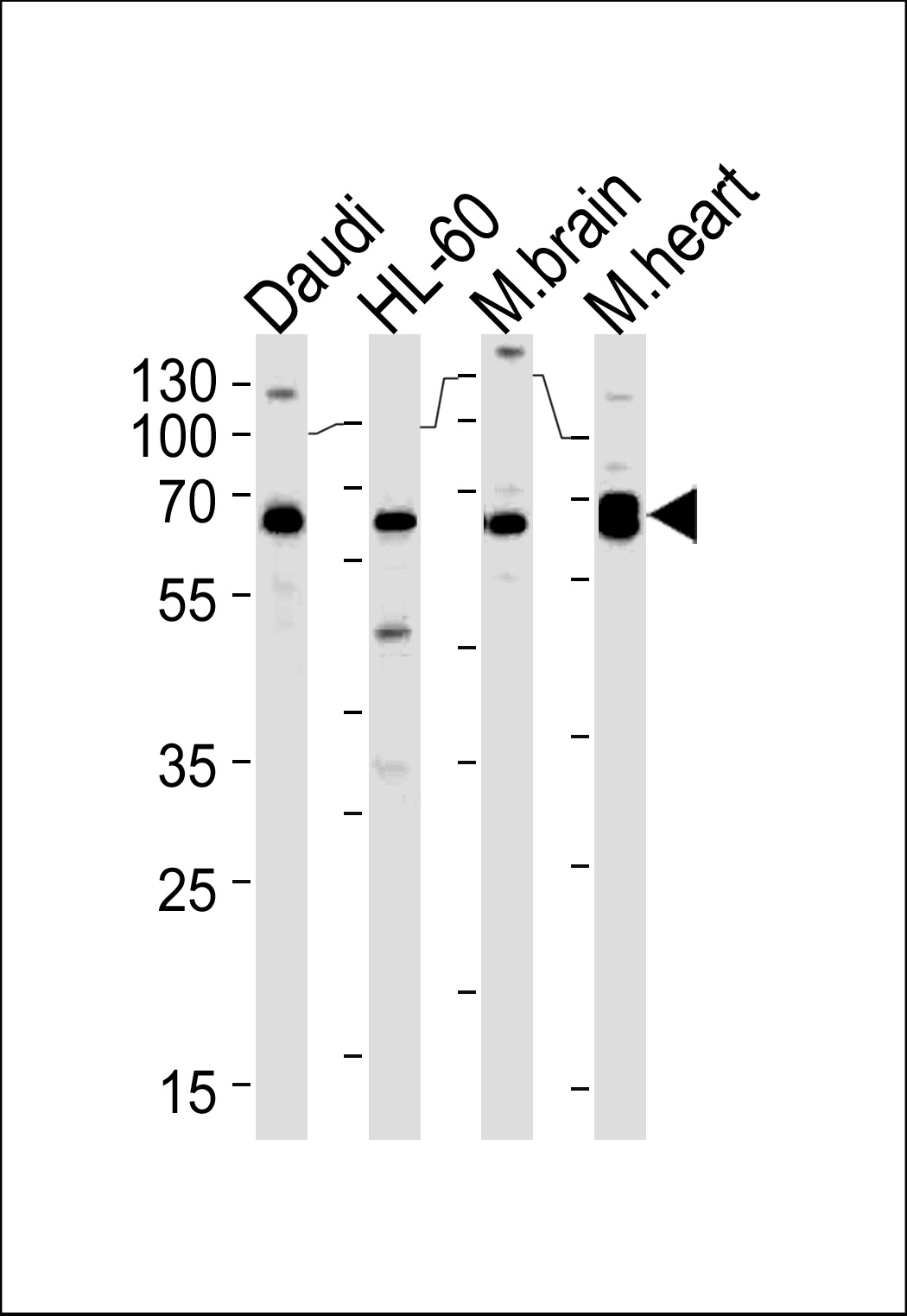
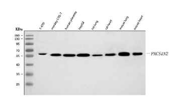
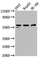
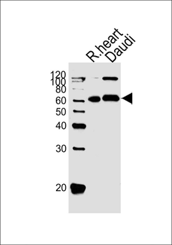
Western blot analysis in Daudi, HL-60 cell line and mouse brain, rat heart lysates (35 ug/lane).This demonstrates the PACSIN2 antibody detected the PACSIN2 protein (arrow).
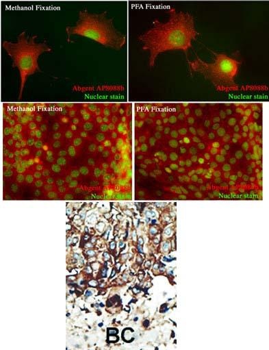
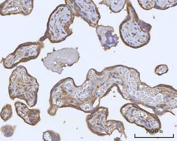
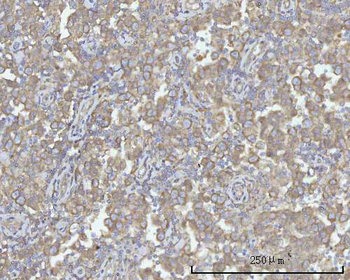
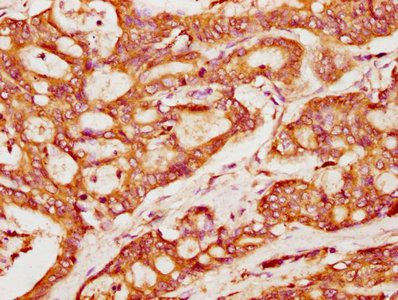
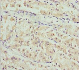
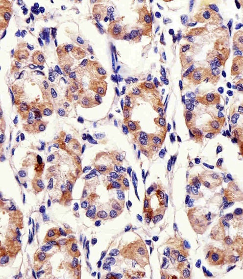
Formalin-fixed and paraffin-embedded human cancer tissue reacted with the primary antibody, which was peroxidase-conjugated to the secondary antibody, followed by DAB staining. BC = breast carcinoma; HC = hepatocarcinoma.
- Item 1 of 9
Anti-PACSIN2 Antibody [orb1676243]
ELISA, FC, ICC, IF, IHC, WB
Human, Monkey, Mouse, Rat
Rabbit
Polyclonal
Unconjugated
10 μg, 100 μg - Item 1 of 4
- Item 1 of 5
PACSIN2 Antibody (C-term) [orb1788460]
IF, IHC-P, WB
Mouse
Human, Rat
Rabbit
Polyclonal
Unconjugated
100 μl - Item 1 of 3
- Item 1 of 3