You have no items in your shopping cart.
Cart summary
Item 1 of 7
Item 1 of 7
Nucleoli Marker Antibody
Catalog Number: orb2638089

| Catalog Number | orb2638089 |
|---|---|
| Category | Antibodies |
| Description | This mAb is part of a panel of reagents which recognizes subcellular organelles or compartments of human cells. These markers may be useful in identification of these organelles in cells, tissues, and biochemical preparations. Clone NM95 antibody recognizes an antigenic marker associated with the nucleoli in human cells. The antibody can be used to stain the nucleoli in cell or tissue preparations and can be used as a marker in subcellular fractions. It produces a speckled pattern in the nuclei of cells of normal and malignant cells. The nucleoli marker antibody can be used with paraformaldehyde fixed frozen tissue or cell preparations and formalin fixed, paraffin-embedded tissue sections. |
| Clonality | Monoclonal |
| Species/Host | Mouse |
| Isotype | Mouse IgG1, kappa |
| Conjugation | Unconjugated |
| Reactivity | Human |
| Buffer/Preservatives | 0.2 mg/ml in 1X PBS with 0.1 mg/ml rAlbumin (US sourced) and 0.05% sodium azide |
| Purity | Protein G affinity chromatography |
| Immunogen | Nuclei of myeloid leukemia biopsy cells were used as the immunogen. |
| Tested applications | FACS, ICC, IF, IHC-P |
| Dilution range | Flow cytometry: 1-2ug/million cells ,Immunofluorescence: 1-2ug/ml,Immunocytochemistry: 1-2ug/ml for 30 min at RT,Immunohistochemistry (FFPE): 1-2ug/ml for 30 min at RT |
| Application notes | The concentration stated for each application is a general starting point. Variations in protocols, secondaries and substrates may require the Nucleoli marker antibody to be titered up or down for optimal performance.1. Staining of formalin-fixed tissues is enhanced by boiling tissue sections in 10mM Citrate Buffer, pH 6.0, for 10-20 min followed by cooling at RT for 20 minutes.2. The prediluted format is supplied in a dropper bottle and is optimized for use in IHC. After epitope retrieval step (if required), drip mAb solution onto the tissue section and incubate at RT for 30 min. |
| Antibody Type | Primary Antibody |
| Clone Number | NM95 |
| Formula | 0.2 mg/ml in 1X PBS with 0.1 mg/ml BSA (US sourced) and 0.05% sodium azide |
| Storage | Maintain refrigerated at 2-8°C for up to 2 weeks. For long term storage store at -20°C in small aliquots to prevent freeze-thaw cycles. |
| Hazard Information | This Nucleoli Marker antibody is available for research use only. |
| Note | For research use only |

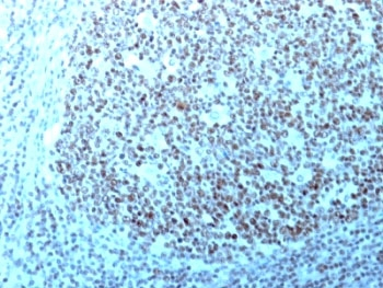
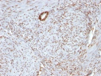
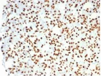
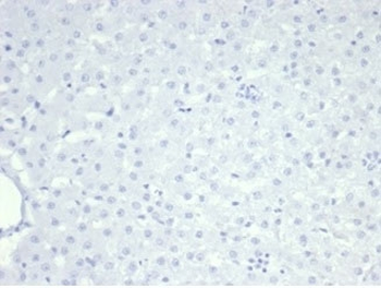
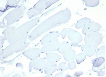
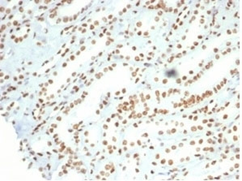
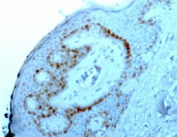
IHC staining of FFPE human skin with Nucleoli Marker antibody. HIER: boil tissue sections in pH6, 10mM citrate buffer, for 10-20 min and allow to cool before testing.

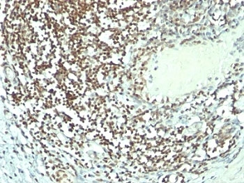
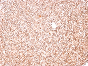
IHC staining of FFPE human tonsil with Nucleoli Marker antibody. HIER: boil tissue sections in pH6, 10mM citrate buffer, for 10-20 min and allow to cool before testing.
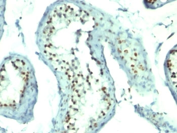
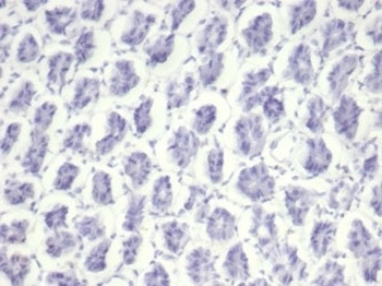
Immunofluorescent staining of FFPE colon carcinoma with Alexa Fluor 488 conjugated Nucleoli Marker antibody. HIER: boil tissue sections in pH6, 10mM citrate buffer, for 10-20 min and allow to cool before testing.

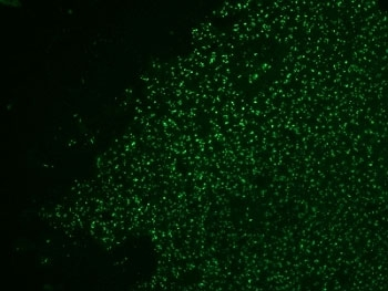
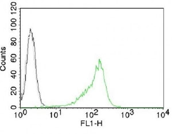
Intracellular FACS testing of 293 cells with Nucleoli Marker antibody (green).

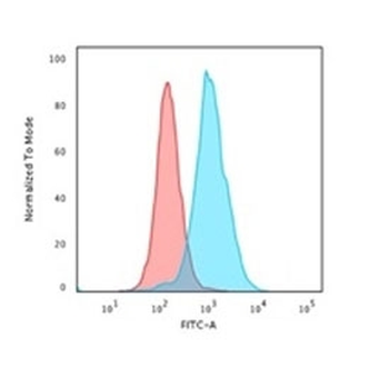
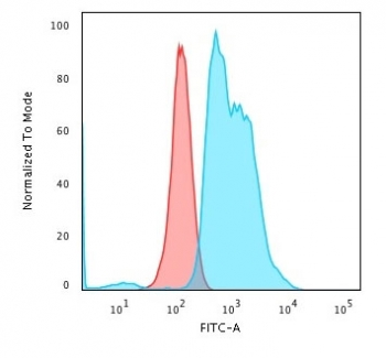
Flow cytometry testing of PFA-fixed human K562 cells with Nucleoli Marker antibody (clone NM95); Red = isotype control, Blue = Nucleoli Marker antibody.
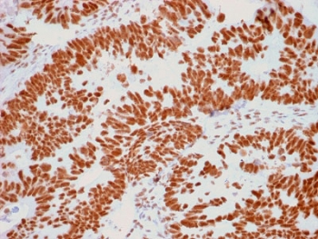
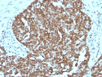
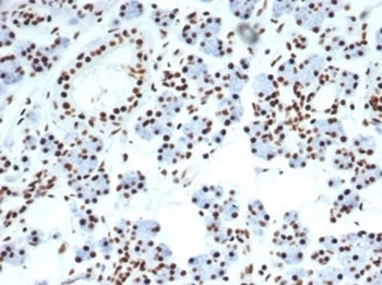
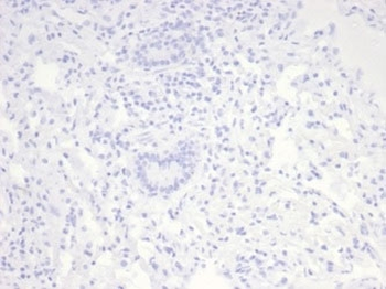
IHC staining of FFPE human colon carcinoma with Nucleoli Marker antibody. HIER: boil tissue sections in pH6, 10mM citrate buffer, for 10-20 min and allow to cool before testing.
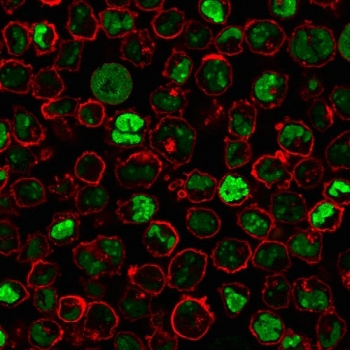
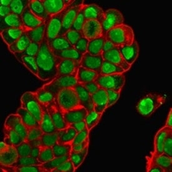
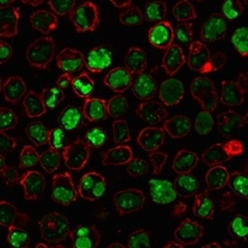
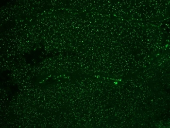
Immunofluorescent staining of PFA-fixed human K562 cells with Nucleoli Marker antibody (green, clone NM95) and Phalloidin (red).
- Item 1 of 9
- Item 1 of 9
- Item 1 of 9
- Item 1 of 9
- Item 1 of 7