You have no items in your shopping cart.
Cart summary
Item 1 of 4
Item 1 of 4
| Catalog Number | orb381911 |
|---|---|
| Category | Antibodies |
| Description | Rabbit polyclonal antibody to NFATC1. |
| Species/Host | Rabbit |
| Clonality | Polyclonal |
| Tested applications | IF, IH, WB |
| Reactivity | Human, Mouse, Rat |
| Immunogen | Recombinant full length protein of human NFAT2 |
| Dilution range | WB: 1:500-2000, IHC-P: 1:50-200, IF/ICC: 1:50-200 |
| Form/Appearance | Liquid in 0.42% Potassium phosphate, 0.87% Sodium chloride, pH 7.3, 30% glycerol, and 0.01% sodium azide. |
| Conjugation | Unconjugated |
| Target | NFATC1 |
| Entrez | 4772 |
| UniProt ID | O88942, O95644 |
| Source | Rabbit |
| Storage | Shipped at 4°C. Upon delivery aliquot and store at -20°C for one year. Avoid freeze/thaw cycles. |
| Buffer/Preservatives | Liquid in 0.42% Potassium phosphate, 0.87% Sodium chloride, pH 7.3, 30% glycerol, and 0.01% sodium azide. |
| Alternative names | NFAT2; NFATC; Nuclear factor of activated T-cells, Read more... |
| Note | For research use only |
| Expiration Date | 12 months from date of receipt. |
Filter by Applications
Filter by Reactivity
Bi, Chun-Sheng et al. Calcitriol inhibits osteoclastogenesis in an inflammatory environment by changing the proportion and function of T helper cell subsets (Th2/Th17) Cell Prolif., 53, e12827 (2020)
Applications
WB
Reactivity
Mouse
Fu, Shengqiang et al. Salidroside promotes osteoblast proliferation and differentiation via the activation of AMPK to inhibit bone resorption of knee osteoarthritis mice Tissue Cell, 79, 101917 (2022)
Applications
WB
Reactivity
Mouse
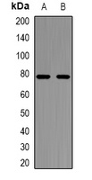
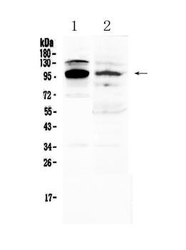
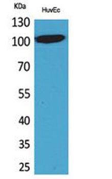
Western blot analysis of NFAT2 expression in Jurkat (A), HL60 (B) whole cell lysates. (Predicted band size: 38; 74-88; 100-101 kD; Observed band size: 74; 101 kD)
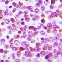
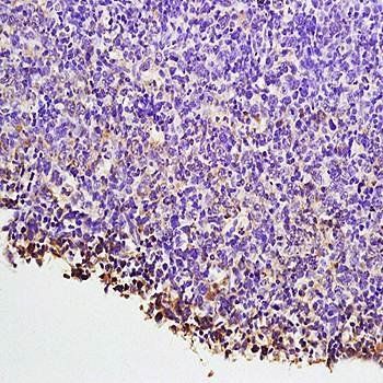
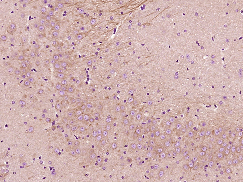
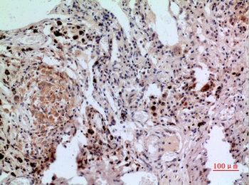
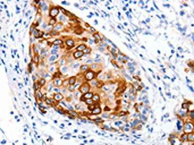
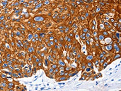
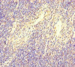
Immunohistochemical analysis of NFAT2 staining in human tonsil formalin fixed paraffin embedded tissue section. The section was pre-treated using heat mediated antigen retrieval with sodium citrate buffer (pH 6.0). The section was then incubated with the antibody at room temperature and detected using an HRP conjugated compact polymer system. DAB was used as the chromogen. The section was then counterstained with haematoxylin and mounted with DPX.
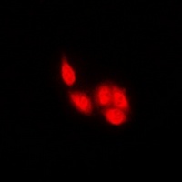
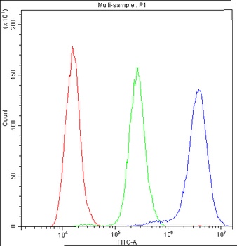
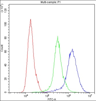
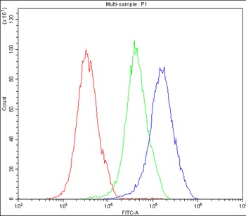
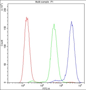
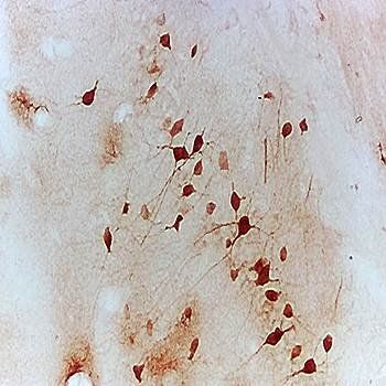
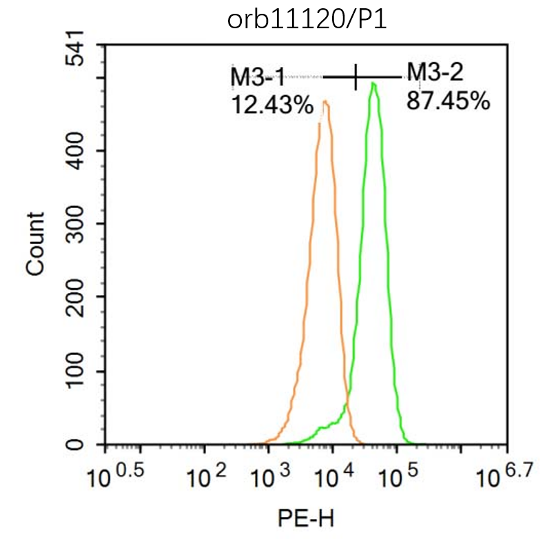
Immunofluorescent analysis of NFAT2 staining in U2OS cells. Formalin-fixed cells were permeabilized with 0.1% Triton X-100 in TBS for 5-10 minutes and blocked with 3% BSA-PBS for 30 minutes at room temperature. Cells were probed with the primary antibody in 3% BSA-PBS and incubated overnight at 4 °C in a humidified chamber. Cells were washed with PBST and incubated with a DyLight 594-conjugated secondary antibody (red) in PBS at room temperature in the dark.
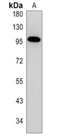
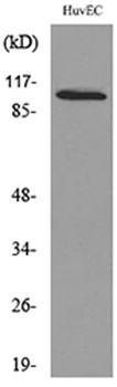
Western blot analysis of NFAT2 expression in mouse thymus (A) whole cell lysates.
- Item 1 of 5
NFAT2/NFATC1 Antibody [orb402344]
ELISA, FC, ICC, IHC, WB
Human, Mouse, Rat
Rabbit
Polyclonal
Unconjugated
10 μg, 100 μg - Item 1 of 4
- Item 1 of 3
- Item 1 of 2
- Item 1 of 1
Submit a review
Filter by Rating

- 5 stars

- 4 stars
- 3 stars
- 2 stars
- 1 stars