You have no items in your shopping cart.
Cart summary
Item 1 of 2
Item 1 of 2
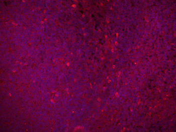
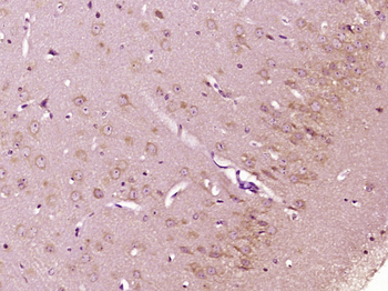
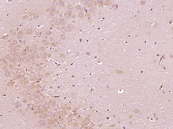
Mitochondrial Marker Antibody
Catalog Number: orb2638081

| Catalog Number | orb2638081 |
|---|---|
| Category | Antibodies |
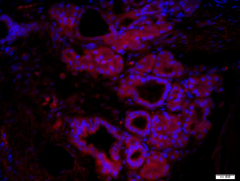
| Description | MAb MTC754 recognizes a 60kDa antigen associated with the mitochondria in cells. It is a part of a new panel of reagents, which recognizes subcellular organelles or compartments of cells. These markers may be useful in identification of these organelles in cells, tissues, and biochemical preparations. MAb MTC754 recognizes an antigen associated with the mitochondria in cells from a wide variety of animals including insects and bacteria. It can be used to stain the mitochondria in cell or tissue preparations and can be used as a mitochondrial marker in subcellular fractions. It produces a spaghetti-like pattern in normal and malignant cells and may be used to stain mitochondria of cells in frozen tissue sections. It can also be used with paraformaldehyde fixed frozen tissue or cell preparations. |
| Species/Host | Mouse |
| Clonality | Monoclonal |
| Clone Number | MTC754 |
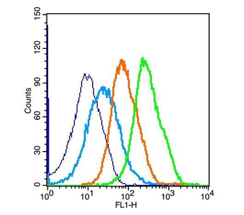
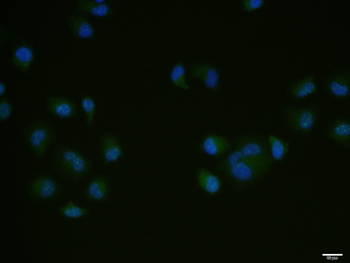
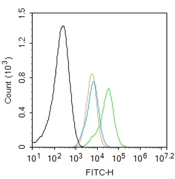
| Tested applications | FACS, IF, IHC-P, WB |
| Reactivity | Human |
| Isotype | Mouse IgG1, kappa |
| Immunogen | The Mitochondrial fraction of HeLa cells was used as the immunogen for this Mitochondria marker antibody. |
| Antibody Type | Primary Antibody |
| Dilution range | Flow cytometry: 0.5-1ug/million cells,Immunofluorescence: 0.5-1ug/ml,Western blot: 0.25-0.5ug/ml,Immunohistochemistry (FFPE): 0.5-1ug/ml for 30 min at RT |
| Purity | Protein G affinity chromatography |
| Conjugation | Unconjugated |
| Formula | 0.2 mg/ml in 1X PBS with 0.1 mg/ml BSA (US sourced) and 0.05% sodium azide |
| Hazard Information | This Mitochondria marker antibody is available for research use only. |
| Storage | Maintain refrigerated at 2-8°C for up to 2 weeks. For long term storage store at -20°C in small aliquots to prevent freeze-thaw cycles. |
| Buffer/Preservatives | 0.2 mg/ml in 1X PBS with 0.1 mg/ml rAlbumin (US sourced) and 0.05% sodium azide |
| Note | For research use only |
| Application notes | The concentration stated for each application is a general starting point. Variations in protocols, secondaries and substrates may require the Mitochondria marker antibody to be titered up or down for optimal performance.1. Staining of FFPE tissues is enhanced by boiling sections in 10mM Tris with 1mM EDTA Buffer, pH 9.0, for 10-20 min followed by cooling at RT for 20 min.2. The prediluted format is supplied in a dropper bottle and is optimized for use in IHC. After epitope retrieval step (if required), drip mAb solution onto the tissue section and incubate at RT for 30 min. |
| Expiration Date | 12 months from date of receipt. |

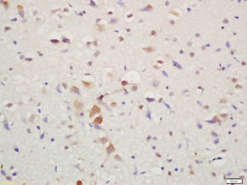
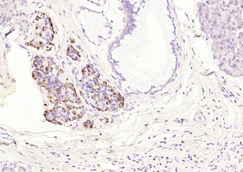
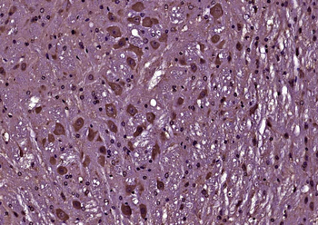
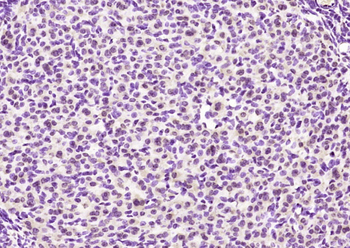
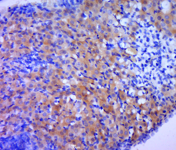
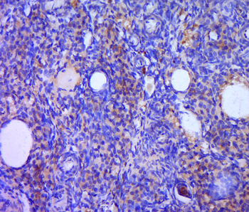
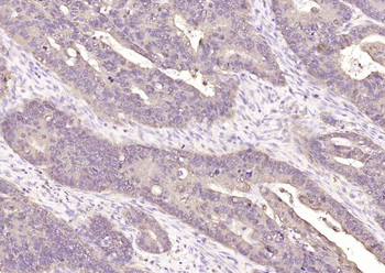
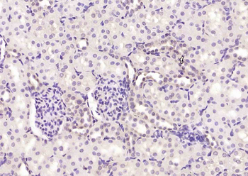
IHC testing of FFPE human renal cell carcinoma with Mitochondria marker antibody
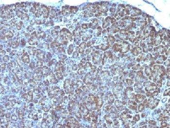
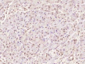
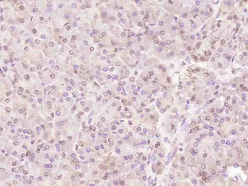
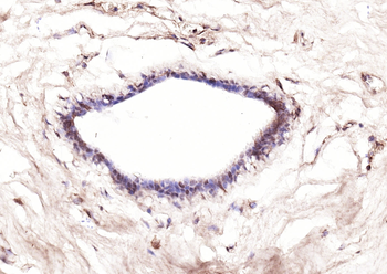
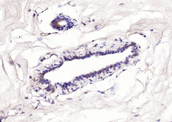
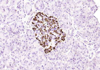
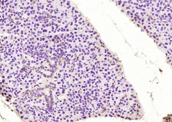
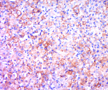
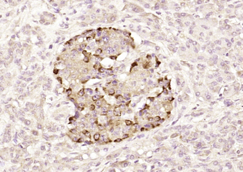
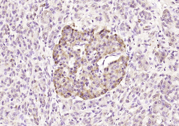
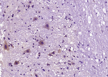
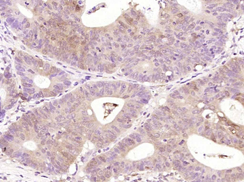
IHC testing of FFPE human pancreas with Mitochondria marker antibody
- Item 1 of 6
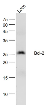
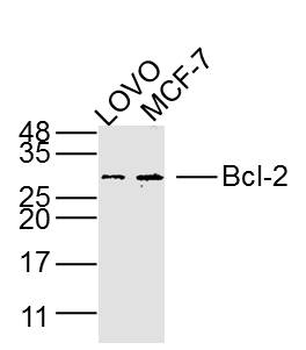
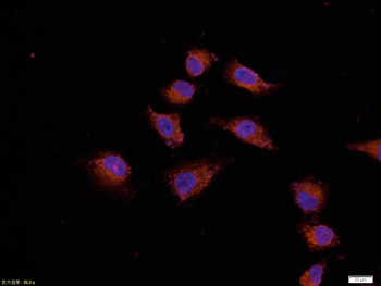
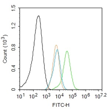
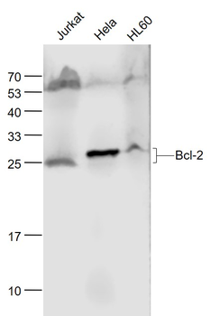
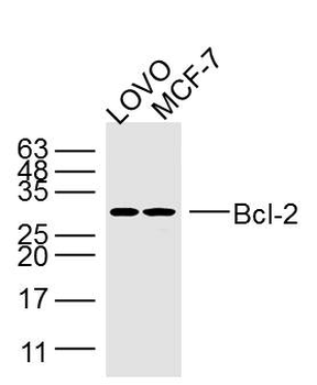
Bcl-2 Rabbit Polyclonal Antibody [orb100697]
ICC, WB
Bovine, Canine, Equine, Porcine, Rabbit
Human, Mouse, Rat
Rabbit
Polyclonal
Unconjugated
50 μl, 100 μl, 200 μl - Item 1 of 9
Bcl-2 Mouse Monoclonal Antibody [orb499778]
ICC, IF, IHC-Fr, IHC-P, WB
Bovine, Canine, Equine, Guinea pig, Mouse, Porcine, Rabbit, Sheep
Human, Rat
Mouse
Monoclonal
Unconjugated
200 μl, 100 μl, 50 μl - Item 1 of 11
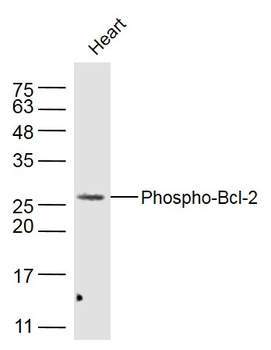
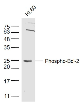
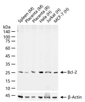
Phospho-Bcl-2 (Thr129) Rabbit Polyclonal Antibody [orb4669]
FC, IF, IHC-Fr, IHC-P, WB
Bovine, Canine, Equine, Porcine, Sheep
Human, Mouse, Rat
Rabbit
Polyclonal
Unconjugated
100 μl, 50 μl, 200 μl - Item 1 of 8
Bcl-2 Rabbit Polyclonal Antibody [orb500830]
ELISA, IF, IHC-Fr, IHC-P, WB
Gallus
Human, Mouse, Rat
Rabbit
Polyclonal
Unconjugated
100 μl, 50 μl - Item 1 of 7
Bcl-2 Mouse Monoclonal Antibody [orb499777]
ICC, IF, IHC-Fr, IHC-P, WB
Bovine, Canine, Equine, Guinea pig, Mouse, Porcine, Rabbit, Sheep
Human, Rat
Mouse
Monoclonal
Unconjugated
200 μl, 50 μl, 100 μl