You have no items in your shopping cart.
Cart summary
Item 1 of 2
Item 1 of 2
Melanoma Antibody / PNL2
Catalog Number: orb2639618

| Catalog Number | orb2639618 |
|---|---|
| Category | Antibodies |
| Description | Anti-PNL2 is a novel melanoma marker monoclonal antibody, which has recently been introduced as an immunohistochemical reagent to stain melanocytes and tumors derived therefrom. The antigen recognized by PNL2 is different from Melan A and gp100. Its epitope is not destroyed by digestion with neuraminidase i.e. its epitope is not glycosylated. Anti-PNL2 may be most useful because of its high sensitivity for metastatic melanoma (87%), as opposed to 76% for anti-HMB45 and 82% for anti-MART-1. Anti-PNL2 labels intra-epidermal nevi while the dermal component of compound nevi are largely non-reactive with anti-PNL2. Antibodies against PNL2, MART-1 (Melan A) and HMB45 stain most clear cell sarcoma cells and a few cells in angiomyolipomas and lymphangioleiomyomatosis. Anti-PNL2 is a useful antibody for the identification of melanomas and clear cell sarcomas. Differential diagnosis is aided by the results from a panel of antibodies, including antibodies against HMB45, MART-1, tyrosinase, and MiTF. |
| Species/Host | Mouse |
| Clonality | Monoclonal |
| Clone Number | PNL2 |
| Tested applications | IF, IHC-P |
| Reactivity | Human |
| Isotype | Mouse IgG1, kappa |
| Immunogen | Melanocyte antigen was used as the immunogen for the Melanoma antibody PNL2. |
| Antibody Type | Primary Antibody |
| Dilution range | Immunofluorescence: 0.5-1ug/ml,Immunohistochemistry (FFPE): 0.5-1ug/ml for 30 min at RT |
| Purity | Protein G affinity chromatography |
| Conjugation | Unconjugated |
| Formula | 0.2 mg/ml in 1X PBS with 0.1 mg/ml BSA (US sourced) and 0.05% sodium azide |
| Hazard Information | This Melanoma antibody PNL2 is available for research use only. |
| UniProt ID | Not Known |
| Storage | Maintain refrigerated at 2-8°C for up to 2 weeks. For long term storage store at -20°C in small aliquots to prevent freeze-thaw cycles. |
| Buffer/Preservatives | 0.2 mg/ml in 1X PBS with 0.1 mg/ml rAlbumin (US sourced) and 0.05% sodium azide |
| Note | For research use only |
| Application notes | Optimal dilution of the pan Cytokeratin antibody cocktail should be determined by the researcher.1. Staining of formalin-fixed tissues requires boiling tissue sections in 10mM Citrate buffer, pH 6.0, for 10-20 min followed by cooling at RT for 20 min.2. The prediluted format is supplied in a dropper bottle and is optimized for use in IHC. After epitope retrieval step (if required), drip mAb solution onto the tissue section and incubate at RT for 30 min. |
| Expiration Date | 12 months from date of receipt. |

IHC anlaylsis of formalin-fixed, paraffin-embedded human melanoma stained with Melanoma antibody (clone PNL2).

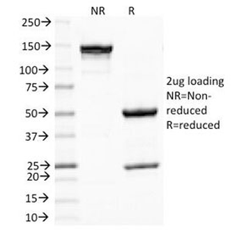
SDS-PAGE Analysis of Purified, BSA-Free Melanoma Antibody (clone PNL2). Confirmation of Integrity and Purity of the Antibody.
- Item 1 of 2
- Item 1 of 2
Melanoma Antibody [PNL2] [orb1252406]
IF, IHC-P, WB
Canine, Human, Mouse
Mouse
Monoclonal
Unconjugated
100 μg

![Melanoma Antibody [PNL2]](/images//pub/media/catalog/product/NewWebsite/15/orb1252406_1.jpg)
![Melanoma Antibody [PNL2]](/images/pub/media/catalog/product/NewWebsite/15/orb1252406_2.jpg)
