You have no items in your shopping cart.
Cart summary
Item 1 of 6
Item 1 of 6
MEK2 Antibody
Catalog Number: orb1262840

| Catalog Number | orb1262840 |
|---|---|
| Category | Antibodies |
| Description | MEK2 Antibody |
| Target | MAP2K2 |
| Clonality | Polyclonal |
| Isotype | Rabbit Ig |
| Conjugation | Unconjugated |
| Reactivity | Human, Mouse |
| Form/Appearance | Liquid |
| Concentration | batch dependent |
| Buffer/Preservatives | Supplied in PBS with 0.09% (W/V) sodium azide. |
| Purification | This antibody is purified through a protein A column, followed by peptide affinity purification. |
| Immunogen | This MEK2 (MAP2K2) antibody is generated from rabbits immunized with a KLH conjugated synthetic peptide between 262-292 amino acids from the Central region of human MEK2 (MAP2K2). |
| UniProt ID | P36507 |
| MW | 44 kDa |
| Tested applications | IF, IHC-P, WB |
| Application notes | For WB starting dilution is: 1:1000For IF starting dilution is: 1:10~50For IHC-P starting dilution is: 1:50~100 |
| Antibody Type | Primary Antibody |
| Storage | Maintain refrigerated at 2-8°C for up to 2 weeks. For long term storage store at -20°C in small aliquots to prevent freeze-thaw cycles. |
| Alternative names | Dual specificity mitogen-activated protein kinase Read more... |
| Note | For research use only |
| NCBI | P36507 |
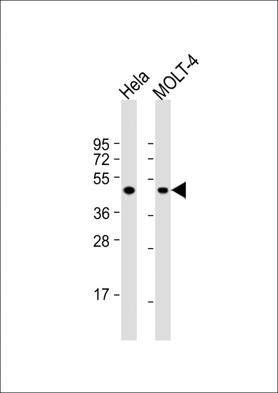
Western Blot at 1:1000 dilution Lane 1: Hela whole cell lysate Lane 2: MOLT-4 whole cell lysate Lysates/proteins at 20 ug per lane.
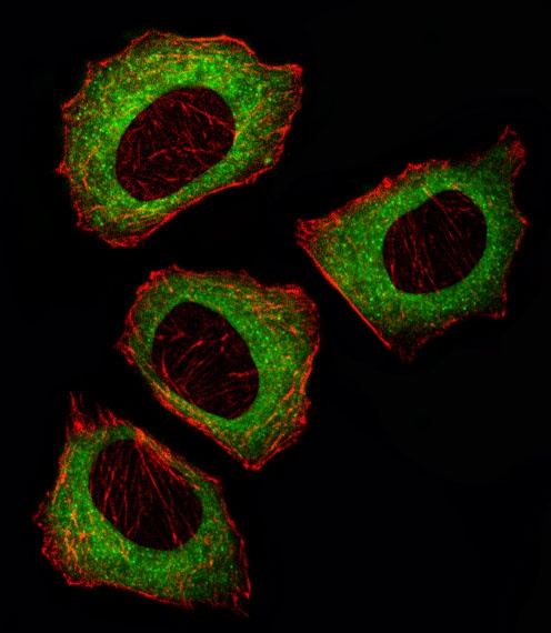
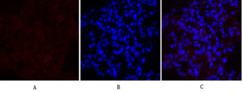
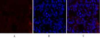
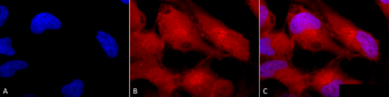
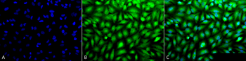
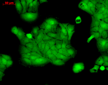
Fluorescent image of U251 cell stained with MEK2 (MAP2K2) Antibody. U251 cells were fixed with 4% PFA (20 min), permeabilized with Triton X-100 (0.1%, 10 min), then incubated with MEK2 primary antibody (1:25). For secondary antibody, Alexa Fluor 488 conjugated donkey anti-rabbit antibody (green) was used (1:400).Cytoplasmic actin was counterstained with Alexa Fluor 555 (red) conjugated Phalloidin (7 units/ml). MEK2 immunoreactivity is localized to Cytoplasm significantly.
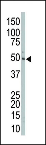
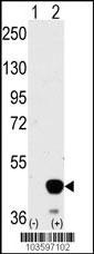
Antibody is used in Western blot to detect MAP2K2 in mouse liver tissue lysate.
Western blot analysis of MAP2K2 using MAP2K2 Antibody.293 cell lysates (2 ug/lane) either nontransfected (Lane 1) or transiently transfected with the MAP2K2 gene (Lane 2).
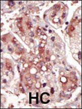
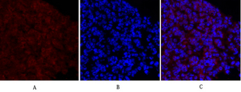
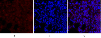
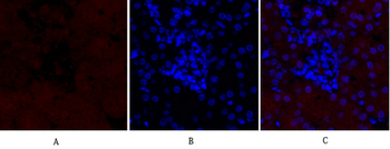
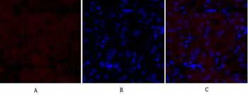
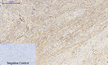
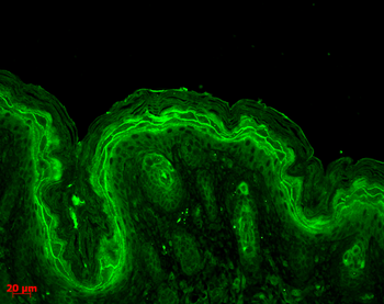
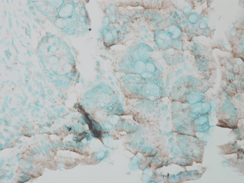
Formalin-fixed and paraffin-embedded human cancer tissue reacted with the primary antibody, which was peroxidase-conjugated to the secondary antibody, followed by DAB staining. BC = breast carcinoma; HC = hepatocarcinoma.
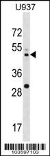
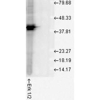
Western blot analysis in U937 cell line lysates (35 ug/lane).
- Item 1 of 11
MEK2 Mouse Monoclonal Antibody [orb499566]
IF, IHC-Fr, IHC-P, WB
Mouse, Rat
Human, Mouse, Rat
Mouse
Monoclonal
Unconjugated
200 μg, 200 μl, 100 μl, 50 μl - Item 1 of 8
Anti-MEK2/MAP2K2 Antibody [orb669099]
ELISA, FC, ICC, IF, IHC, WB
Human, Mouse, Rat
Rabbit
Polyclonal
Unconjugated
100 μg, 10 μg - Item 1 of 7
MEK-1/2 Polyclonal Antibody [orb1413122]
IF, IHC-P, WB
Human, Mouse, Rat
Rabbit
Polyclonal
Unconjugated
100 μl - Item 1 of 6
Erk1/2 Antibody: APC [orb151495]
ICC, IF, IHC
Bovine, Drosophila, Frog, Gallus, Human, Mouse, Rat, Sheep
Rabbit
Polyclonal
APC
100 μl - Item 1 of 6
Erk1/2 Antibody: Biotin [orb151496]
ELISA, ICC, IF, IHC, WB
Bovine, Drosophila, Frog, Gallus, Human, Mouse, Rat, Sheep
Rabbit
Polyclonal
Biotin
100 μl