You have no items in your shopping cart.
Cart summary
Item 1 of 4
Item 1 of 4
MAP2 Antibody (Ascites)
Catalog Number: orb652061

| Catalog Number | orb652061 |
|---|---|
| Category | Antibodies |
| Description | Mouse monoclonal antibody to MAP2 |
| Target | MAP2 |
| Clonality | Monoclonal |
| Species/Host | Mouse |
| Isotype | IgG1,K |
| Conjugation | Unconjugated |
| Reactivity | Human |
| Form/Appearance | Mouse monoclonal antibody supplied in crude ascites with 0.09% (W/V) sodium azide. |
| Immunogen | Recombinant Protein |
| UniProt ID | P11137 |
| MW | 199526 |
| Tested applications | IF, IHC-P, WB |
| Dilution range | WB: 1:1000 |
| Antibody Type | Primary Antibody |
| Clone Number | 159CT34.12.3.4 |
| Storage | Maintain refrigerated at 2-8°C for up to 2 weeks. For long term storage store at -20°C in small aliquots to prevent freeze-thaw cycles |
| Alternative names | Anti-Microtubule-associated protein 2 antibody, an Read more... |
| Note | For research use only |
| NCBI | NP_001034627.1, NP_114035.2, NP_114033.2, NP_002365.3 |
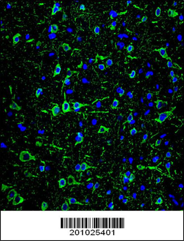
Confocal immunofluorescent analysis of MAP2 Antibody with brain tissue followed by Alexa Fluor 488-conjugated goat anti-mouse lgG (green). DAPI was used to stain the cell nuclear (blue).
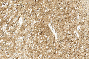
Immunohistochemical analysis of paraffin-embedded Human brain section using Pink1. diluted at 1:500 dilution. A undiluted biotinylated goat polyvalent antibody was used as the secondary, followed by DAB staining.
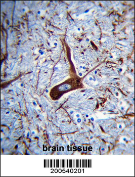
MAP2 Antibody (Ascites) immunohistochemistry analysis in formalin fixed and paraffin embedded human brain tissue followed by peroxidase conjugation of the secondary antibody and DAB staining. This data demonstrates the use of MAP2 Antibody (Ascites) for immunohistochemistry. Clinical relevance has not been evaluated.
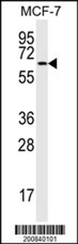
MAP2 Antibody western blot analysis in MCF-7 cell line lysates (15 μg/lane). This demonstrates the MAP2 antibody detected the MAP2 protein (arrow).

MAP2 monoclonal antibody [orb1677294]
IF, IHC, WB
Human
Mouse
Monoclonal
Unconjugated
200 μl, 100 μl, 50 μl
