You have no items in your shopping cart.
Cart summary
Item 1 of 4
Item 1 of 4
LRP12 Antibody
Catalog Number: orb1269992

| Catalog Number | orb1269992 |
|---|---|
| Category | Antibodies |
| Description | LRP12 Antibody |
| Target | LRP12 |
| Clonality | Polyclonal |
| Isotype | Rabbit Ig |
| Conjugation | Unconjugated |
| Reactivity | Human |
| Predicted Reactivity | Monkey, Mouse |
| Form/Appearance | Liquid |
| Concentration | batch dependent |
| Buffer/Preservatives | Supplied in PBS with 0.09% (W/V) sodium azide. |
| Purification | This antibody is purified through a protein A column, followed by peptide affinity purification. |
| Immunogen | This LRP12 antibody is generated from rabbits immunized with a KLH conjugated synthetic peptide between 635-662 amino acids from the C-terminal region of human LRP12. |
| UniProt ID | Q9Y561 |
| MW | 95 kDa |
| Tested applications | FC, IF, IHC-P, WB |
| Application notes | For WB starting dilution is: 1:1000For IHC-P starting dilution is: 1:10~50For IF starting dilution is: 1:10~50For FACS starting dilution is: 1:10~50 |
| Antibody Type | Primary Antibody |
| Storage | Maintain refrigerated at 2-8°C for up to 2 weeks. For long term storage store at -20°C in small aliquots to prevent freeze-thaw cycles. |
| Alternative names | Low-density lipoprotein receptor-related protein 1 Read more... |
| Note | For research use only |
| NCBI | Q9Y561 |

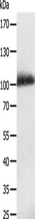
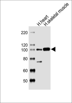
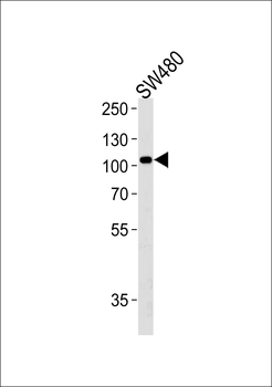
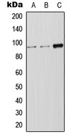
Western blot analysis of lysate from SW480 cell line, using LRP12 Antibody at 1:1000 at each lane
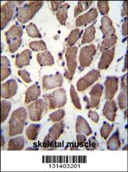
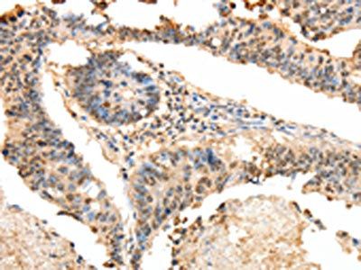
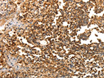
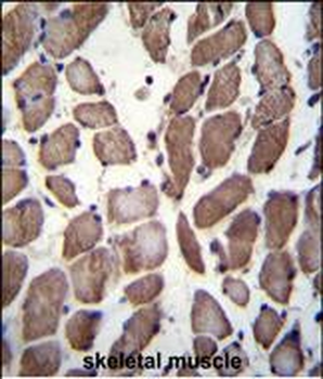
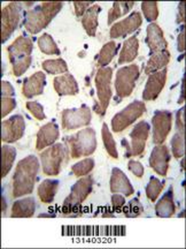
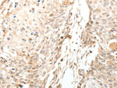
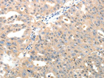
LRP12 Antibody immunohistochemistry analysis in formalin fixed and paraffin embedded human skeletal muscle followed by peroxidase conjugation of the secondary antibody and DAB staining.
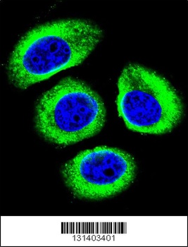
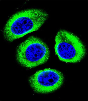
Confocal immunofluorescent analysis of LRP12 Antibody with U-251MG cell followed by Alexa Fluor 488-conjugated goat anti-rabbit lgG (green). DAPI was used to stain the cell nuclear (blue).
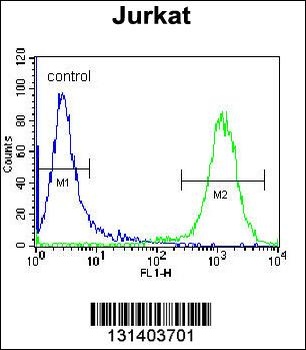
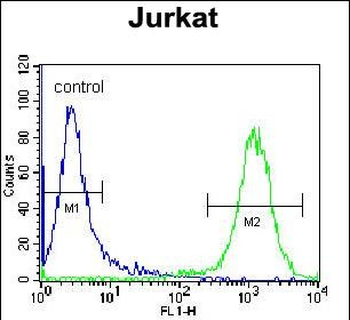
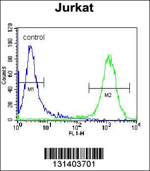
Flow cytometric analysis of Jurkat cells (right histogram) compared to a negative control cell (left histogram). FITC-conjugated donkey-anti-rabbit secondary antibodies were used for the analysis.
- Item 1 of 3
- Item 1 of 4
- Item 1 of 4
LRP12 Antibody (C-term) [orb29733]
FC, IF, IHC-P, WB
Mouse
Human
Rabbit
Polyclonal
Unconjugated
100 μl, 50 μl - Item 1 of 2
- Item 1 of 1