You have no items in your shopping cart.
Cart summary
Item 1 of 4
Item 1 of 4
Ki67/Mki67 Antibody
Catalog Number: orb763060
| Catalog Number | orb763060 |
|---|---|
| Category | Antibodies |
| Description | Ki67/Mki67 Antibody |
| Species/Host | Rabbit |
| Clonality | Polyclonal |
| Tested applications | ELISA, IHC |
| Reactivity | Human, Mouse, Rat |
| Isotype | Rabbit IgG |
| Immunogen | E.coli-derived mouse Mki67 recombinant protein (Position: F29-S3177). |
| Concentration | Adding 0.2 ml of distilled water will yield a concentration of 500 μg/ml. |
| Dilution range | Immunohistochemistry (Paraffin-embedded Section), 2-5μg/ml, Human, Mouse, Rat Direct ELISA, 0.1-0.5μg/ml, Mouse |
| Form/Appearance | Lyophilized |
| Conjugation | Unconjugated |
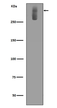
| MW | 358 kDa |
| UniProt ID | E9PVX6 |
| Storage | Store at -20˚C for one year from date of receipt. After reconstitution, at 4˚C for one month. It can also be aliquotted and stored frozen at -20˚C for six months. Avoid repeated freeze-thaw cycles. |
| Note | For research use only |
| Application notes | Tested Species: In-house tested species with positive results. Other applications have not been tested. Optimal dilutions should be determined by end users. Add 0.2ml of distilled water will yield a concentration of 500ug/ml. |
| Expiration Date | 12 months from date of receipt. |
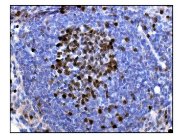
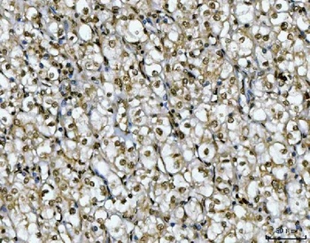
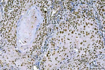
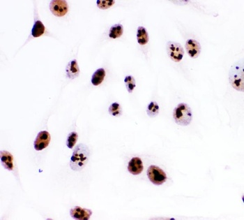
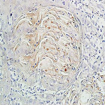
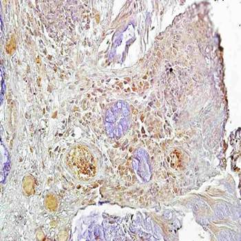
IHC analysis of Ki67/Mki67 using anti-Ki67/Mki67 antibody (orb763060). Ki67/Mki67 was detected in a paraffin-embedded section of human tonsil tissue. Heat mediated antigen retrieval was performed in EDTA buffer (pH 8.0, epitope retrieval solution). The tissue section was blocked with 10% goat serum. The tissue section was then incubated with 2 μg/ml rabbit anti-Ki67/Mki67 Antibody (orb763060) overnight at 4°C. Biotinylated goat anti-rabbit IgG was used as secondary antibody and incubated for 30 minutes at 37°C. The tissue section was developed using Strepavidin-Biotin-Complex (SABC) (Catalog # orb90444) with DAB as the chromogen.
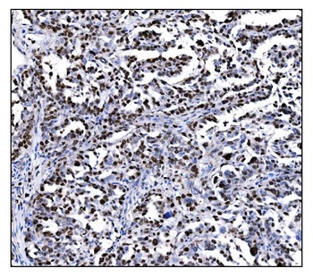
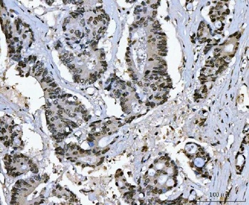
IHC analysis of Ki67/Mki67 using anti-Ki67/Mki67 antibody (orb763060). Ki67/Mki67 was detected in a paraffin-embedded section of human ovarian cancer tissue. Heat-mediated antigen retrieval was performed in EDTA buffer (pH 8.0, epitope retrieval solution). The tissue section was blocked with 10% goat serum. The tissue section was then incubated with 2 μg/ml rabbit anti-Ki67/Mki67 Antibody (orb763060) overnight at 4°C. Biotinylated goat anti-rabbit IgG was used as secondary antibody and incubated for 30 minutes at 37°C. The tissue section was developed using Strepavidin-Biotin-Complex (SABC) (Catalog # orb90444) with DAB as the chromogen.
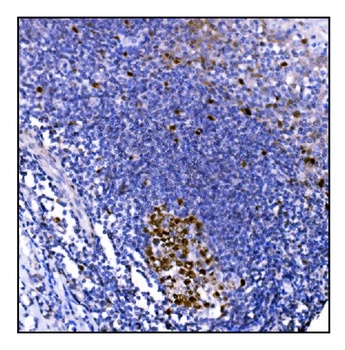
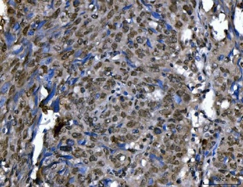
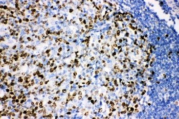
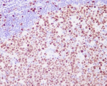
IHC analysis of Ki67/Mki67 using anti-Ki67/Mki67 antibody (orb763060). Ki67/Mki67 was detected in a paraffin-embedded section of mouse lymphaden tissue. Heat mediated antigen retrieval was performed in EDTA buffer (pH 8.0, epitope retrieval solution). The tissue section was blocked with 10% goat serum. The tissue section was then incubated with 2 μg/ml rabbit anti-Ki67/Mki67 Antibody (orb763060) overnight at 4°C. Biotinylated goat anti-rabbit IgG was used as secondary antibody and incubated for 30 minutes at 37°C. The tissue section was developed using Strepavidin-Biotin-Complex (SABC) (Catalog # orb90444) with DAB as the chromogen.
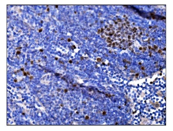
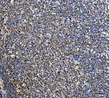
IHC analysis of Ki67/Mki67 using anti-Ki67/Mki67 antibody (orb763060). Ki67/Mki67 was detected in a paraffin-embedded section of rat lymphaden tissue. Heat mediated antigen retrieval was performed in EDTA buffer (pH 8.0, epitope retrieval solution). The tissue section was blocked with 10% goat serum. The tissue section was then incubated with 2 μg/ml rabbit anti-Ki67/Mki67 Antibody (orb763060) overnight at 4°C. Biotinylated goat anti-rabbit IgG was used as secondary antibody and incubated for 30 minutes at 37°C. The tissue section was developed using Strepavidin-Biotin-Complex (SABC) (Catalog # orb90444) with DAB as the chromogen.
- Item 1 of 5
- Item 1 of 4
- Item 1 of 2
- Item 1 of 2
Ki67 MKI67 Rabbit Monoclonal Antibody [orb547195]
FC, ICC, IF, IHC, WB
Human
Rabbit
Monoclonal
Unconjugated
30 μl, 100 μl - Item 1 of 2
Submit a review
Filter by Rating

- 5 stars

- 4 stars
- 3 stars
- 2 stars
- 1 stars