You have no items in your shopping cart.
Cart summary
Item 1 of 4
Item 1 of 4
IRAK2 Antibody
Catalog Number: orb1238892
| Catalog Number | orb1238892 |
|---|---|
| Category | Antibodies |
| Description | IRAK2 Antibody |
| Species/Host | Rabbit |
| Clonality | Polyclonal |
| Tested applications | ELISA, ICC, IF, WB |
| Predicted Reactivity | Mouse |
| Reactivity | Human |
| Isotype | IgG |
| Immunogen | IRAK2 antibody was raised against a peptide corresponding to amino acids near the carboxy terminus of human IRAK2.The immunogen is located within amino acids 530 - 580 of IRAK-2. |
| Concentration | 1 mg/ml |
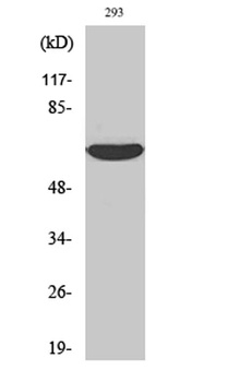
| Dilution range | IRAK-2 antibody can be used for detection of IRAK2 by Western blot at 2 μg/mL. A 65 kDa band should be detected. This polyclonal antibody can also detect IRAK2 by immunocytochemistry at 2 μg/mL. For immunofluorescence start at 10 μg/mL.Antibody validated: Western Blot in human samples; Immunocytochemistry in human samples and Immunofluorescence in human samples. All other applications and species not yet tested. |
| Form/Appearance | Liquid |
| Conjugation | Unconjugated |
| MW | 65 kDa |
| Target | IRAK2 |
| UniProt ID | O43187 |
| NCBI | AF026273 |
| Storage | IRAK-2 antibody can be stored at 4°C for three months and -20°C, stable for up to one year. As with all antibodies care should be taken to avoid repeated freeze thaw cycles. Antibodies should not be exposed to prolonged high temperatures. |
| Buffer/Preservatives | IRAK-2 Antibody is supplied in PBS containing 0.02% sodium azide. |
| Alternative names | IRAK-2 Antibody: IRAK-2, Interleukin-1 receptor-as Read more... |
| Note | For research use only |
| Application notes | IRAK-2 antibody can be used for detection of IRAK2 by Western blot at 2 μg/mL. A 65 kDa band should be detected. This polyclonal antibody can also detect IRAK2 by immunocytochemistry at 2 μg/mL. For immunofluorescence start at 10 μg/mL.Antibody validated: Western Blot in human samples; Immunocytochemistry in human samples and Immunofluorescence in human samples. All other applications and species not yet tested. |
| Expiration Date | 12 months from date of receipt. |
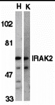
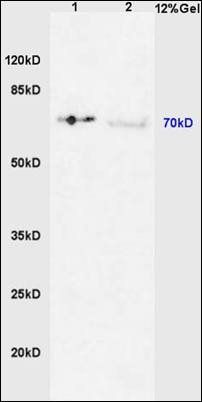
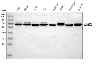
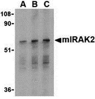
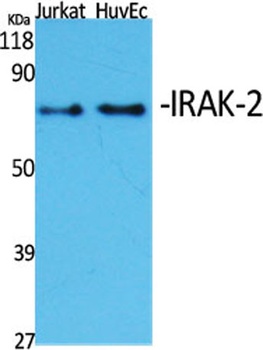
Western blot analysis of IRAK2 in HeLa (H) and K562 (K) whole cell lysate with IRAK2 antibody (C2) at 2 µg/mL.
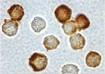
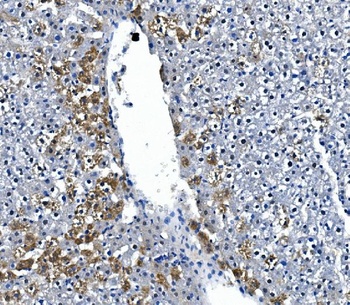
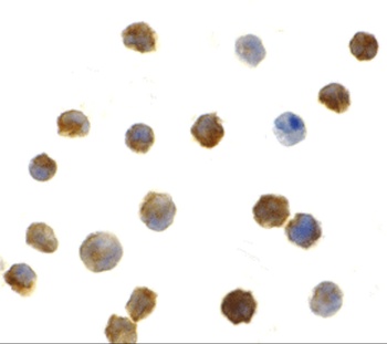
Immunocytochemical staining of HeLa cells using IRAK2 antibody at 2 µg/mL.
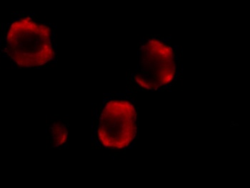
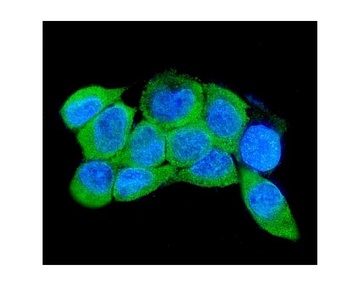
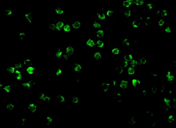
Immunofluorescence of IRAK-2 in Hela cells with IRAK-2 antibody at 10 µg/mL.
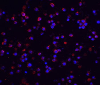
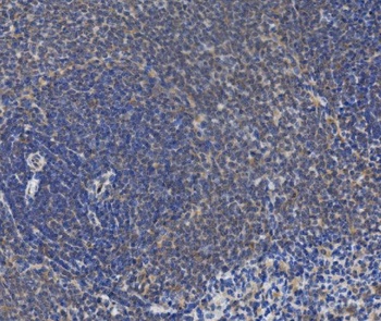
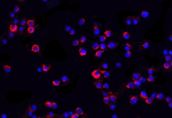
Immunofluorescence of IRAK-M in human spleen tissue with IRAK M antibody at 20 µg/mL. Green: IRAK-2 Antibody (orb1238892) Blue: DAPI staining.
- Item 1 of 6
IRAK1 (phospho-Thr387) antibody [orb6223]
FC, IHC-P, WB
Bovine, Canine, Porcine, Rabbit
Human, Mouse, Rat
Rabbit
Polyclonal
Unconjugated
200 μl, 50 μl, 100 μl - Item 1 of 5
- Item 1 of 4
Irak2 Antibody [orb1238891]
ELISA, ICC, IF, WB
Bovine, Rat
Human, Mouse
Rabbit
Polyclonal
Unconjugated
0.1 mg, 0.02 mg - Item 1 of 3
- Item 1 of 2
Submit a review
Filter by Rating

- 5 stars

- 4 stars
- 3 stars
- 2 stars
- 1 stars