You have no items in your shopping cart.
Cart summary
Item 1 of 2
Item 1 of 2
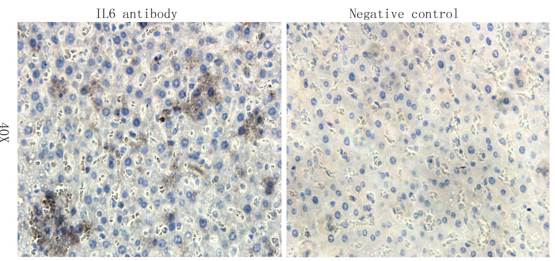
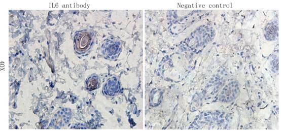
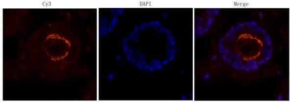
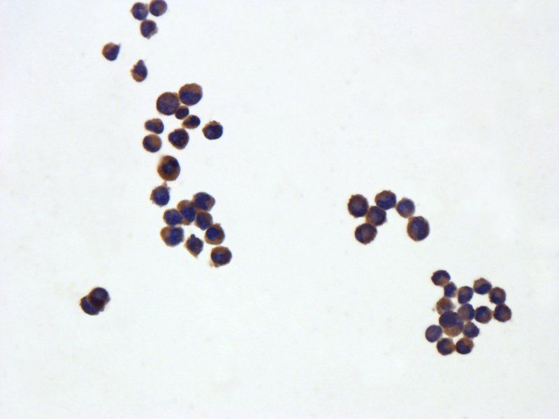
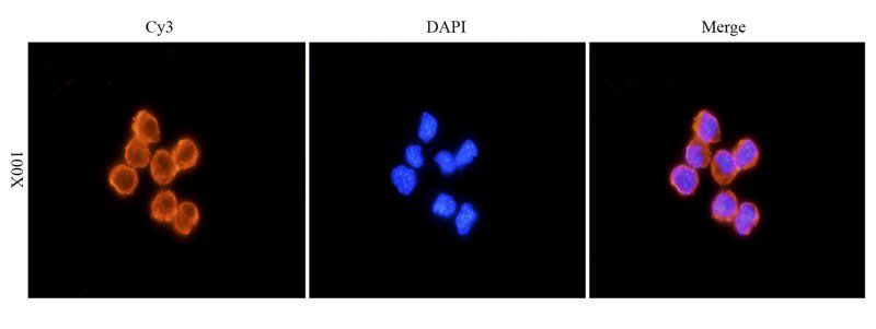
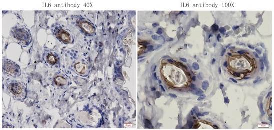
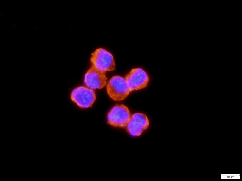
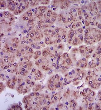
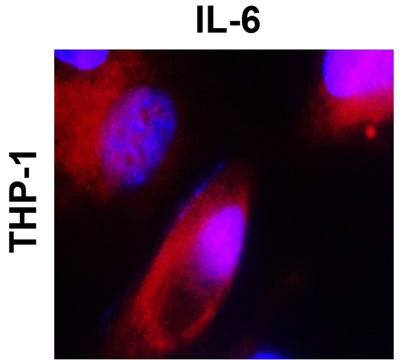
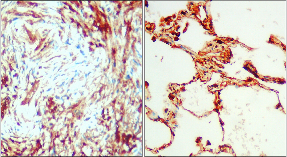
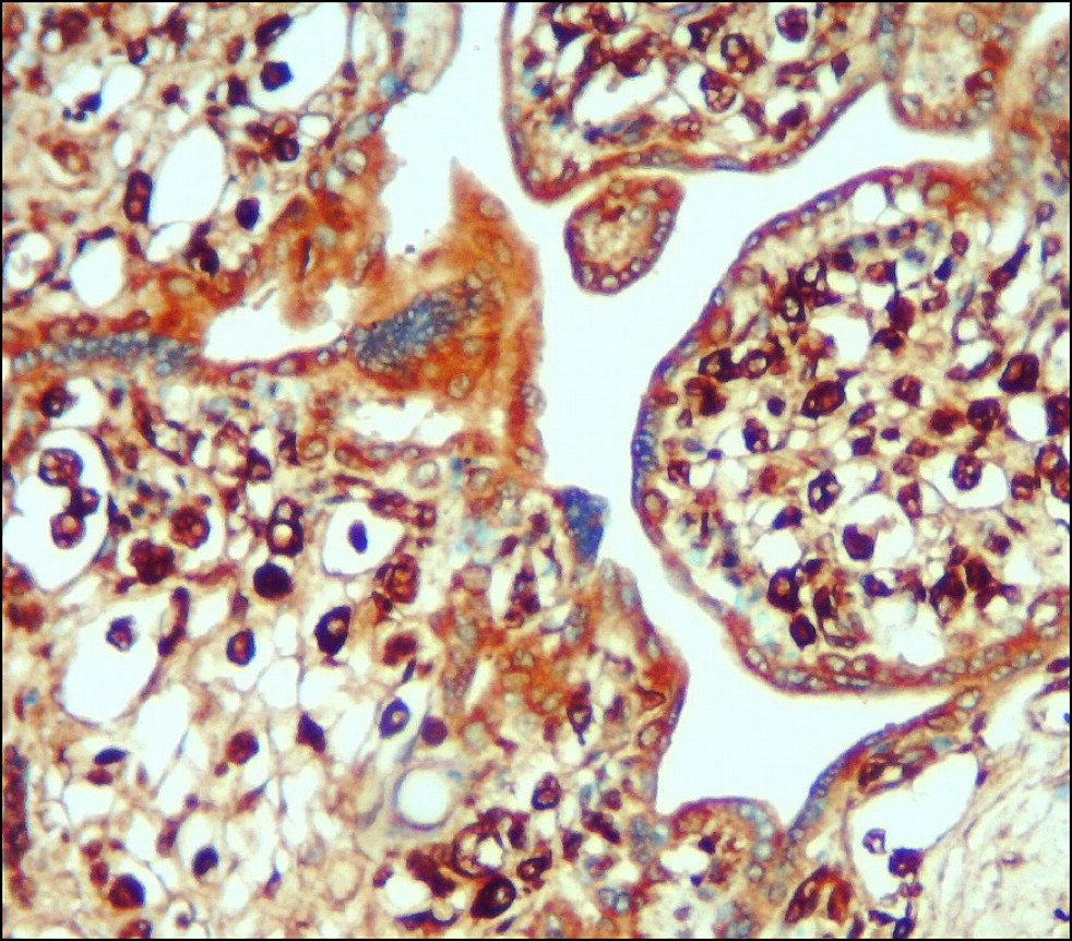
IL6 antibody
Catalog Number: orb750682

| Catalog Number | orb750682 |
|---|---|
| Category | Antibodies |
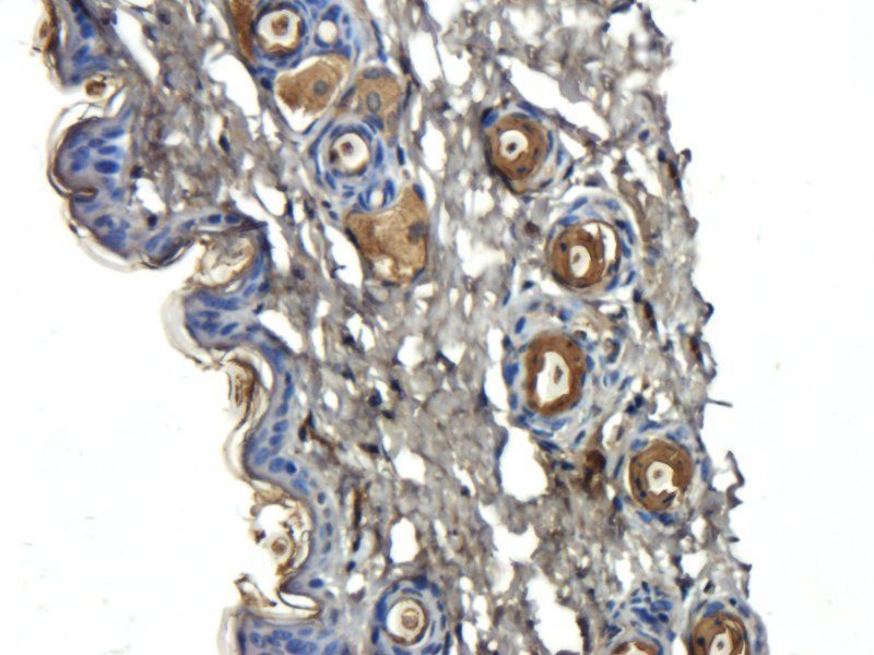
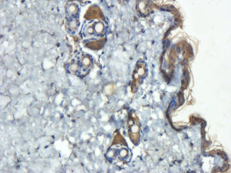
| Description | IL6 antibody |
| Species/Host | Rabbit |
| Clonality | Polyclonal |
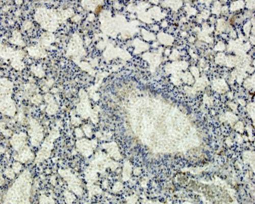
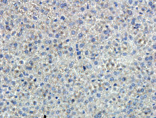
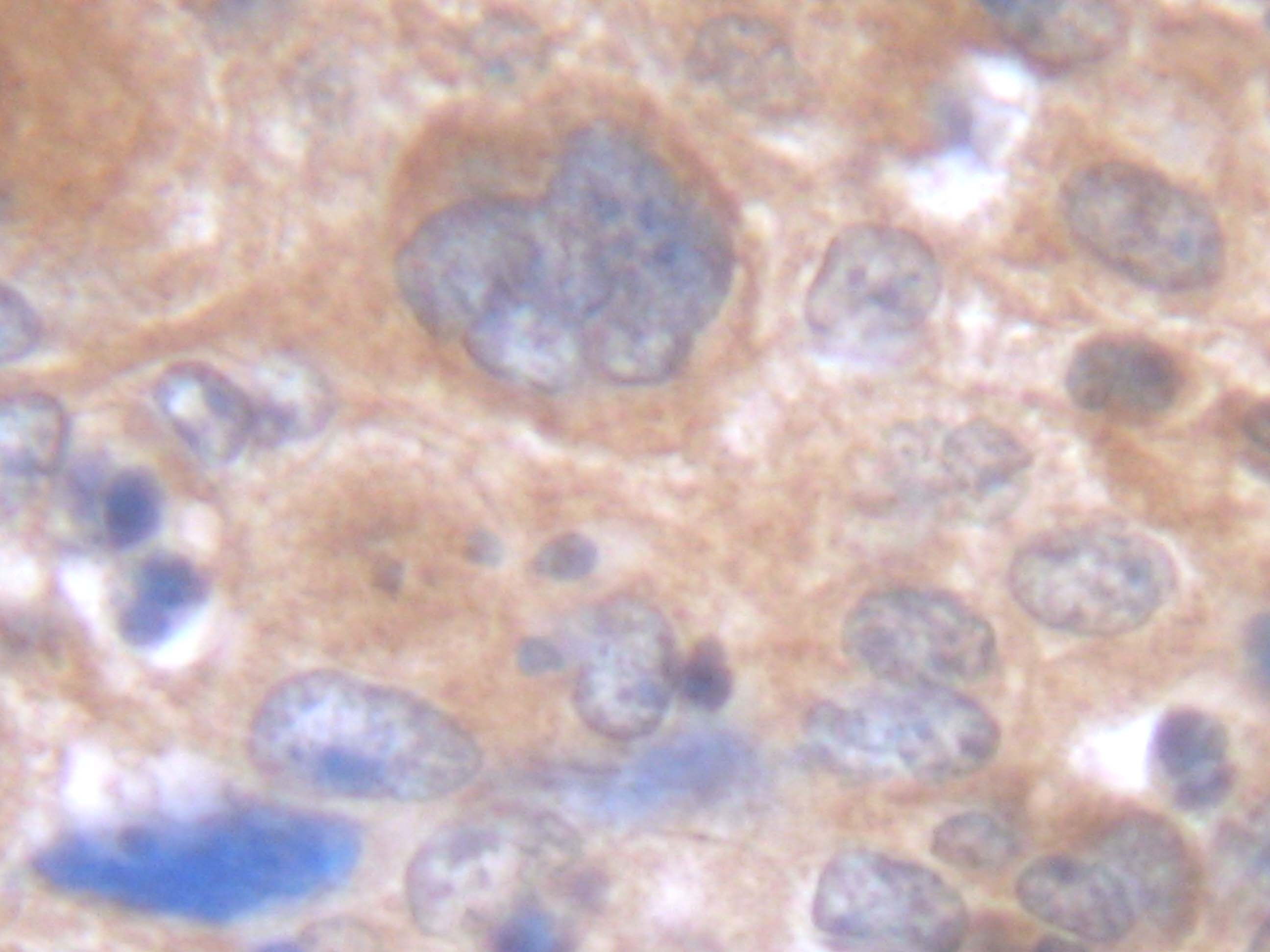
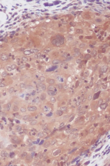
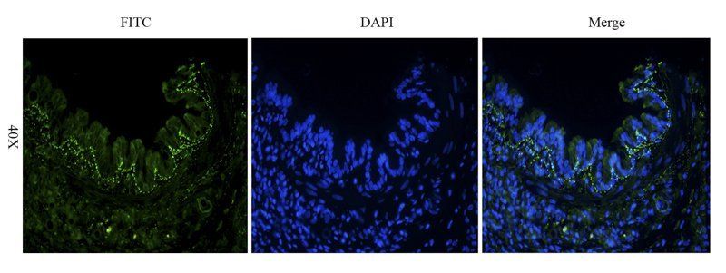
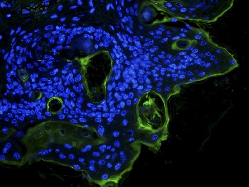
| Tested applications | DOT, ELISA, IHC, IP, WB |
| Reactivity | Human |
| Isotype | Antiserum |
| Immunogen | This whole rabbit serum was prepared by repeated immunizations with recombinant human IL-6 produced in E.coli. |
| Concentration | 80 mg/mL |
| Dilution range | ELISA: 1:1,000 - 1:5,000, IHC: 1:400 - 1:800, IP: 1:400 - 1:800, WB: 1:500 - 1:2,000 |
| Form/Appearance | Liquid (sterile filtered) |
| Purity | Anti-IL-6 antiserum detects recombinant and native human IL-6 present in body fluids and cell supernatants in various assays (ie. IL-1 stimulated IL-6 production from fibroblasts). This product has minimal reactivity with mouse IL-6. |
| Conjugation | Unconjugated |
| UniProt ID | P05231 |
| NCBI | NP_000591.1 |
| Storage | Store vial at -20° C or below prior to opening. This vial contains a relatively low volume of reagent (25 µL). To minimize loss of volume dilute 1:10 by adding 225 µL of the buffer stated above directly to the vial. Recap, mix thoroughly and briefly centrifuge to collect the volume at the bottom of the vial. Use this intermediate dilution when calculating final dilutions as recommended below. Store the vial at -20°C or below after dilution. Avoid cycles of freezing and thawing. |
| Buffer/Preservatives | None |
| Alternative names | HSF, Hybridoma growth factor, HGF, Hybridoma plasm Read more... |
| Note | For research use only |
| Application notes | IL-6 antibody has been tested in western blot. This antibody is suitable for use in neutralizations, ELISA, radioimmunoassay, and immunoprecipitation. Reactivity in other immunoassays is unknown. Expect ~23.7kDa. In Western blot analysis of natural cell products or human body fluids, multiple bands of IL-6 will appear due to the variable amount of glycosylation on the molecule. For immunoblot use the supernatants or lysates of 2 x 10⁶ endotoxin-stimulated human peripheral blood mononuclear cells (PBMC). PBMC are stimulated for 24 hours with 1% (v/v) human serum plus 10ng/mL E. coli LPS. For immunoprecipitation, pre-clearing with a non-specific rabbit IgG is helpful to reduce background. The antiserum is useful for neutralization of human of IL-6 activity in bioassays. For neutralization, incubate the sample with a 1:400 dilution of the antiserum for at least 4 hours before being tested. A control of similarly diluted normal rabbit IgG (heat inactivated) is recommended. In neutralization experiments in vitro, this antibody does not result in enhanced activity of IL-6. However, because antibodies to IL-6 may act as a soluble receptor in vivo, some antibodies to IL-6 act as carriers and enhance IL-6 activity. |
| Expiration Date | 12 months from date of receipt. |

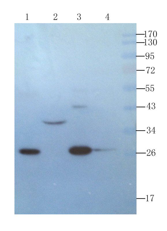
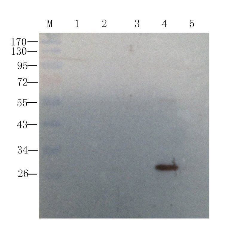
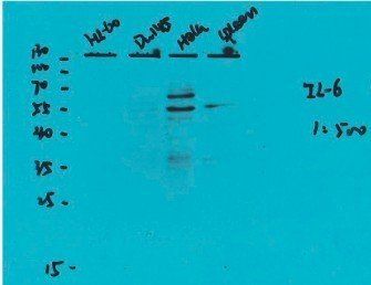
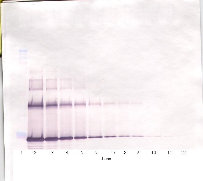
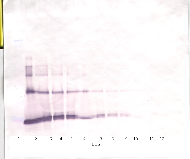
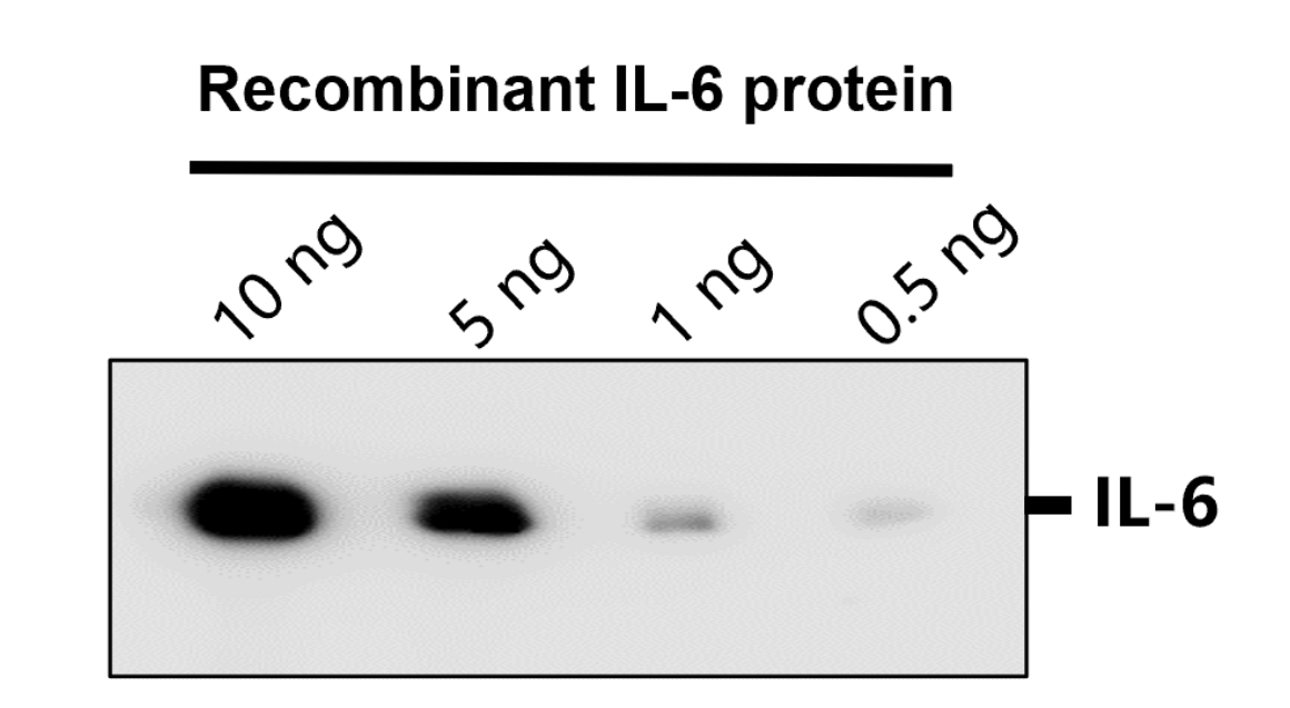
Western Blot of Rabbit Anti-IL-6 Antibody. Lane 1: Opal Prestained Molecular Weight Marker. Lane 2: Human Rec. IL-6/HeLa Whole Cell Lysate [0.05 µg/10 µg]. Lane 3: Human Rec. IL-6/HeLa Whole Cell Lysate [0.02 µg/10 µg]. Lane 4: Human Rec. IL-6/HeLa Whole Cell Lysate [0.01 µg/10 µg]. Lane 5: HeLa Whole Cell Lysate (p/n orb348668) [10 µg]. Primary Antibody: Anti-IL-6 at 1:1000 overnight at 2-8°C. Secondary Antibody: Goat Anti-Rabbit IgG HRP conjugate (p/n orb347654) at 1:70000 for 30 mins at RT. Block: BlockOut Buffer (p/n orb348644). Predicted MW: ~23 kDa. Observed MW: ~19 Exposure: 2 seconds.
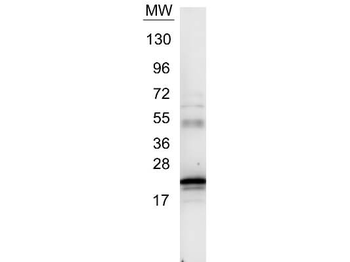
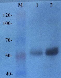
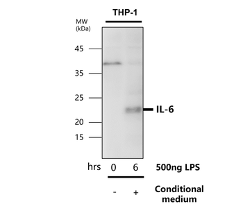
Western blot using Biorbyt's Anti-IL6 antibody. Protein was resolved on a 4-20% Tris-Glycine gel by SDS-PAGE and transferred onto nitrocellulose. The blot shows detection of a band ~21 kDa in size corresponding to anti-IL6 antibody. Molecular weight markers are also shown (MW). After transfer, the membrane was blocked for 30 minutes with 1% BSA-TBST. Detection occurred using peroxidase conjugated Goat anti-Rabbit IgG (p/n orb347654) secondary antibody diluted 1:40000 in blocking buffer (p/n orb348637) for 30 min at RT followed by reaction with FemtoMax™ chemiluminescent substrate.
- Item 1 of 17
- Item 1 of 7
- Item 1 of 5
- Item 1 of 4
- Item 1 of 5
Submit a review
Filter by Rating

- 5 stars

- 4 stars
- 3 stars
- 2 stars
- 1 stars
