You have no items in your shopping cart.
Cart summary
Item 1 of 5
Item 1 of 5
IGF2BP1/IMP1
Catalog Number: orb20564

| Catalog Number | orb20564 |
|---|---|
| Category | Antibodies |
| Description | Goat polyclonal antibody to IGF2BP1 |
| Target | IMP1 / ZBP1 |
| Clonality | Polyclonal |
| Species/Host | Goat |
| Conjugation | Unconjugated |
| Reactivity | Bovine, Canine, Human, Mouse, Rat |
| Buffer/Preservatives | Supplied at 0.5 mg/ml in Tris saline, 0.02% sodium azide, pH 7.3 with 0.5% bovine serum albumin. Aliquot and store at -20°C. Minimize freezing and thawing. |
| Purification | Purified from goat serum by ammonium sulphate precipitation followed by antigen affinity chromatography using the immunizing peptide. |
| Protein Sequence | EKVFAEHKISYSGQ |
| RRID | AB_10753702 |
| MW | 63.5; 48.6 |
| Tested applications | ELISA, FC, IF, IHC, WB |
| Dilution range | ELISA: 1:16000, WB: 0.3-1 μg/ml, IHC-P: 3.75 μg/ml |
| Application notes | ELISA: Peptide ELISA: antibody detection limit dilution 1:16000.WB: Approx 65kDa band observed in lysates of cell line K562 (calculated MW of 63.5kDa according to NP_006537.3;). Recommended concentration: 0.3-1 μg/ml. |
| Storage | Aliquot and store at -20°C. Minimize freezing and thawing. |
| Alternative names | anti coding region determinant-binding protein ant Read more... |
| Note | For research use only |
| Entrez | 10642 |
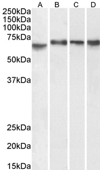
0.1 µg/ml staining of Caco-2 (A), (0.3 µg/ml) K562 (B), nuclear HepG2 (C) and (1 µg/ml) nuclear NIH3T3 (D) cell lysate (35 µg protein in RIPA buffer). Detected by chemiluminescence.
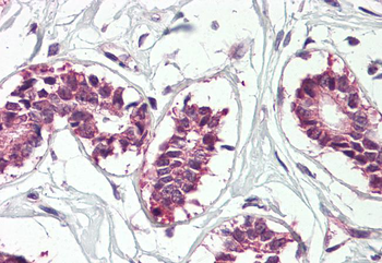
3.75 µg/ml staining of paraffin embedded Human Breast. Steamed antigen retrieval with citrate buffer pH 6, AP-staining.
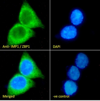
Immunofluorescence analysis of paraformaldehyde fixed HepG2 cells, permeabilized with 0.15% Triton. Primary incubation 1hr (10 ug/ml) followed by Alexa Fluor 488 secondary antibody (2 ug/ml), showing cytoplasmic staining. The nuclear stain is DAPI (blue). Negative control: Unimmunized goat IgG (10 ug/ml) followed by Alexa Fluor 488 secondary antibody (2 ug/ml).
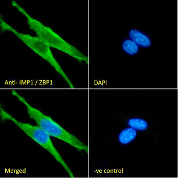
Immunofluorescence analysis of paraformaldehyde fixed NIH3T3 cells, permeabilized with 0.15% Triton. Primary incubation 1hr (10 ug/ml) followed by Alexa Fluor 488 secondary antibody (2 ug/ml), showing nuclear and cytoplasmic staining. The nuclear stain is DAPI (blue). Negative control: Unimmunized goat IgG (10 ug/ml) followed by Alexa Fluor 488 secondary antibody (2 ug/ml).
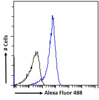
Flow cytometric analysis of paraformaldehyde fixed HepG2 cells (blue line), permeabilized with 0.5% Triton. Primary incubation 1hr (10 ug/ml) followed by Alexa Fluor 488 secondary antibody (1 ug/ml). IgG control: Unimmunized goat IgG (black line) followed by Alexa Fluor 488 secondary antibody.

IGF2BP1/IMP1 polyclonal antibody [orb646677]
IF, IHC, WB
Human, Mouse, Rat
Rabbit
Polyclonal
Unconjugated
200 μl, 100 μl, 50 μl[KO Validated] IGF2BP1/IMP1 polyclonal antibody [orb1718557]
WB
Human, Mouse, Rat
Rabbit
Polyclonal
Unconjugated
100 μl, 50 μl
