You have no items in your shopping cart.
Cart summary
Item 1 of 12
Item 1 of 12
IDH2 Antibody
Catalog Number: orb312128

| Catalog Number | orb312128 |
|---|---|
| Category | Antibodies |
| Description | IDH2 Antibody |
| Species/Host | Rabbit |
| Clonality | Polyclonal |
| Tested applications | FC, ICC, IF, IHC, IHC-Fr, WB |
| Predicted Reactivity | Hamster |
| Reactivity | Human, Mouse, Rat |
| Isotype | Rabbit IgG |
| Immunogen | A synthetic peptide corresponding to a sequence at the C-terminus of human IDH2 (413-447aa KDLAGCIHGLSNVKLNEHFLNTTDFLDTIKSNLDR), identical to the related mouse and rat sequences. |
| Concentration | Adding 0.2 ml of distilled water will yield a concentration of 500 μg/ml. |
| Form/Appearance | Lyophilized |
| Conjugation | Unconjugated |
| MW | 50909 MW |
| UniProt ID | P48735 |
| Storage | Store at -20˚C for one year from date of receipt. After reconstitution, at 4˚C for one month. It can also be aliquotted and stored frozen at -20˚C for six months. Avoid repeated freeze-thaw cycles. |
| Alternative names | Isocitrate dehydrogenase [NADP], mitochondrial;IDH Read more... |
| Note | For research use only |
| Application notes | Tested Species: In-house tested species with positive results. By Heat: Boiling the paraffin sections in 10mM citrate buffer, pH6.0, for 20mins is required for the staining of formalin/paraffin sections. Other applications have not been tested. Optimal dilutions should be determined by end users. . Add 0.2ml of distilled water will yield a concentration of 500ug/ml. |
| Expiration Date | 12 months from date of receipt. |
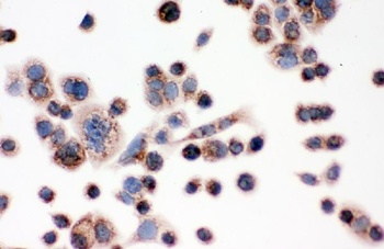
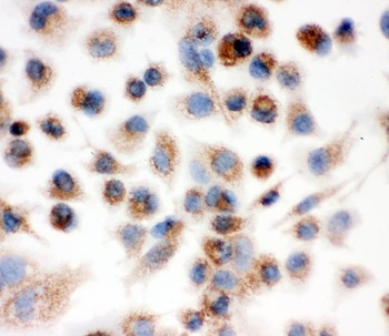
ICC analysis of IDH2 using anti-IDH2 antibody.IDH2 was detected in immunocytochemical section of SW480 cell.Biotinylated goat anti-rabbit IgG was used as secondary antibody.
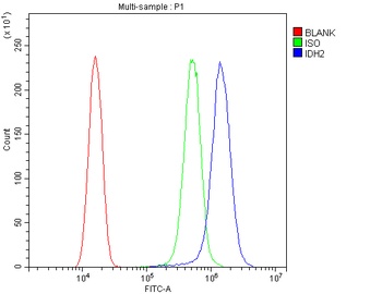
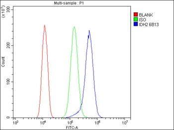
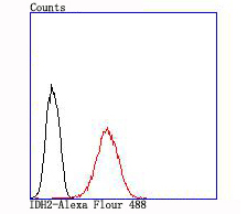
Flow Cytometry analysis of SiHa cells using anti-IDH2 antibody (Blue line).Isotype control antibody (Green line) was rabbit IgG .Unlabelled sample (Red line) was also used as a control.
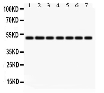
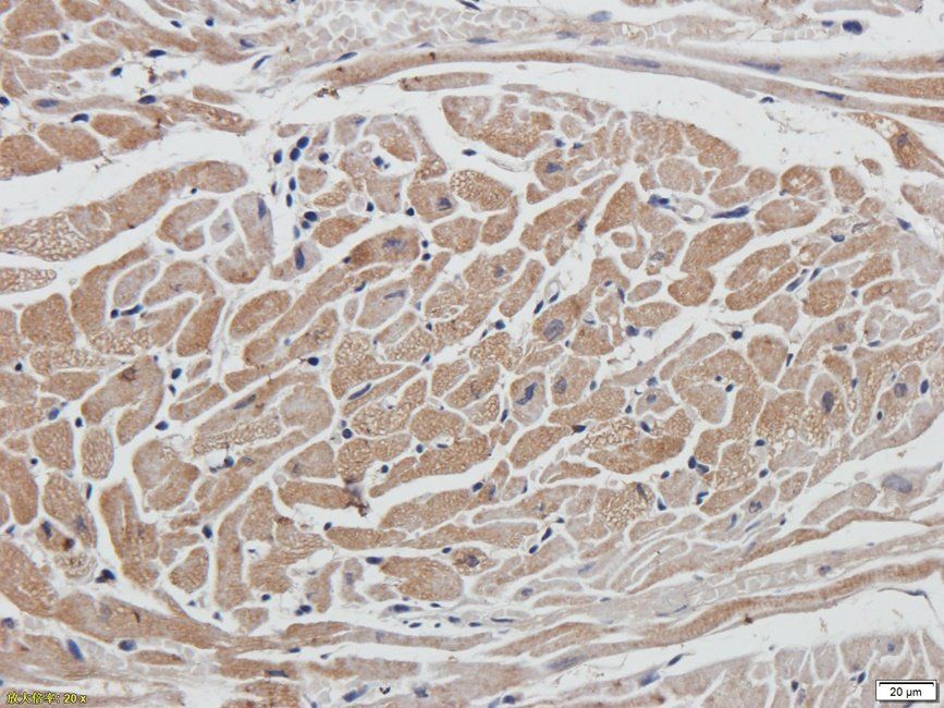
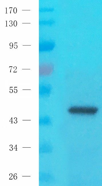
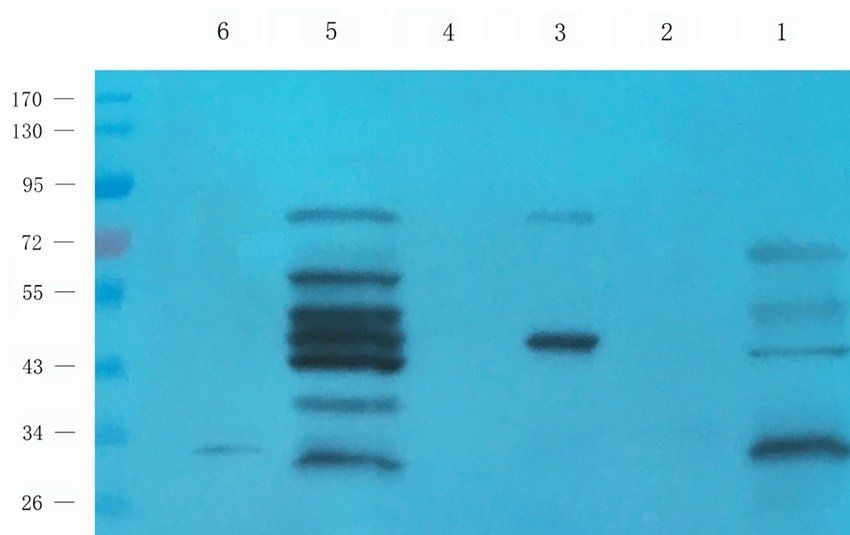
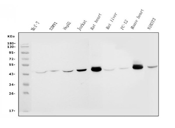
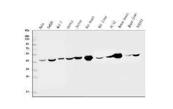
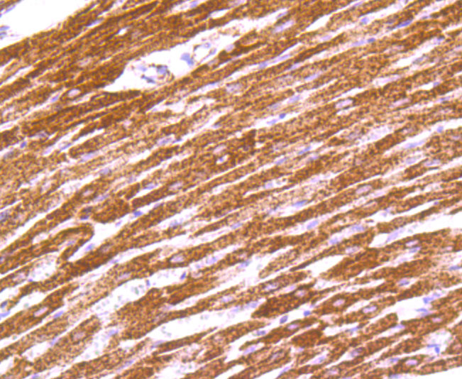
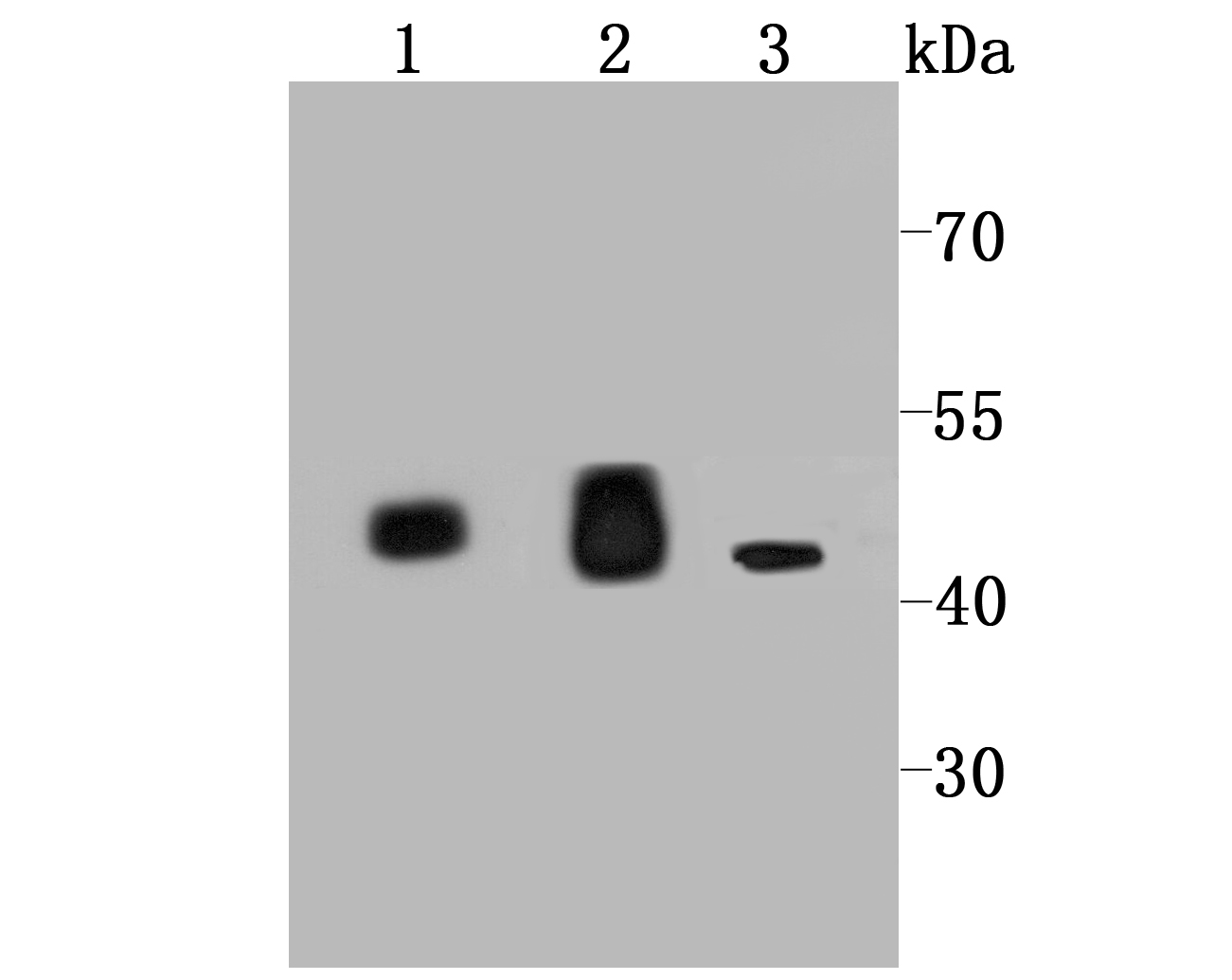
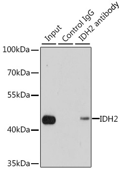
WB analysis of IDH2 using anti-IDH2 antibody.Lane 1:Rat Cardiac Muscle Tissue;2:Rat Liver Tissue;3:NIH3T3 Cell;4:SW620 Cell;5:HELA Cell;6:MCF-7 Cell;7:22RV1 Cell.
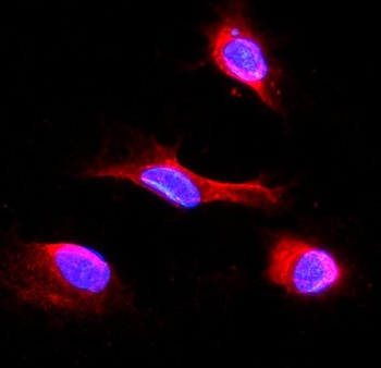
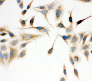
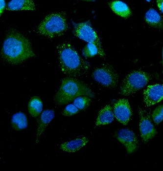
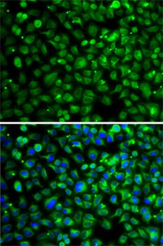
IF analysis of IDH2 using anti-IDH2 antibody. IDH2 was detected in immunocytochemical section of NRK cells.
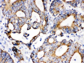
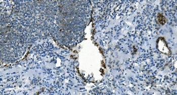
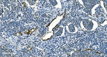
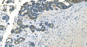
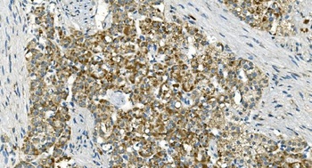
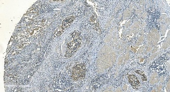
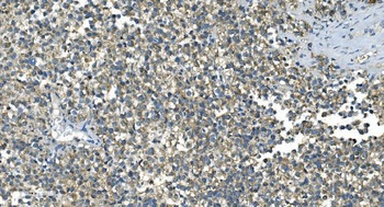
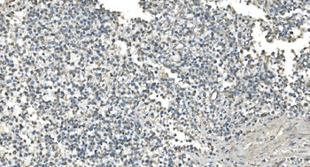
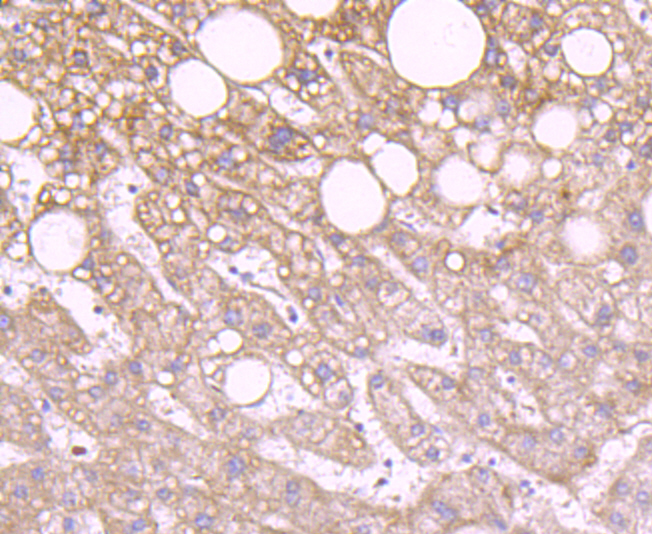
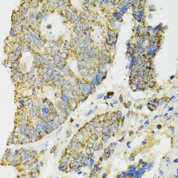
IHC analysis of IDH2 using anti-IDH2 antibody. IDH2 was detected in paraffin-embedded section of Human Intestinal Cancer Tissue.
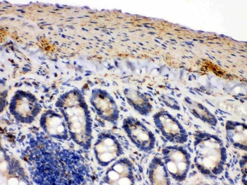
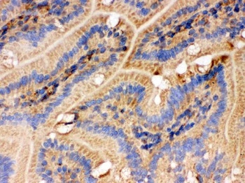
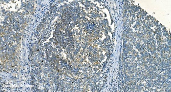
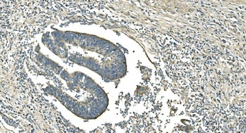
IHC analysis of IDH2 using anti-IDH2 antibody. IDH2 was detected in paraffin-embedded section of Mouse Intestine Tissue.
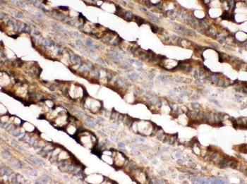
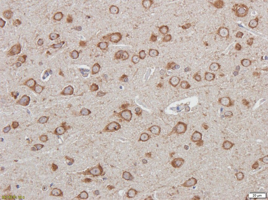
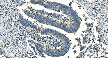
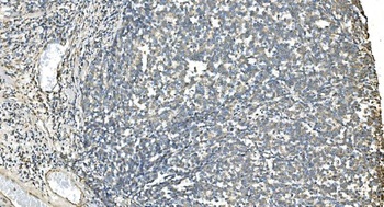
IHC analysis of IDH2 using anti-IDH2 antibody. IDH2 was detected in paraffin-embedded section of Rat Intestine Tissue.
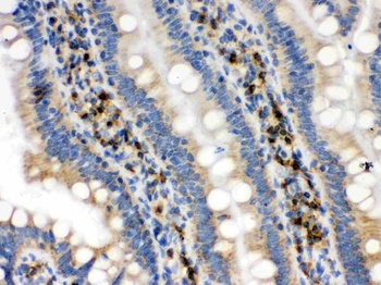
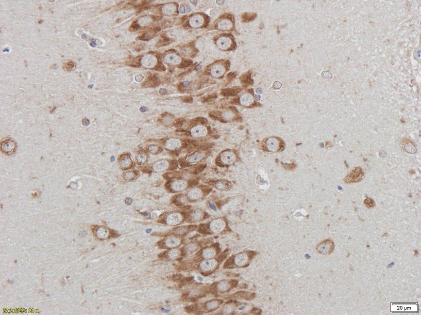
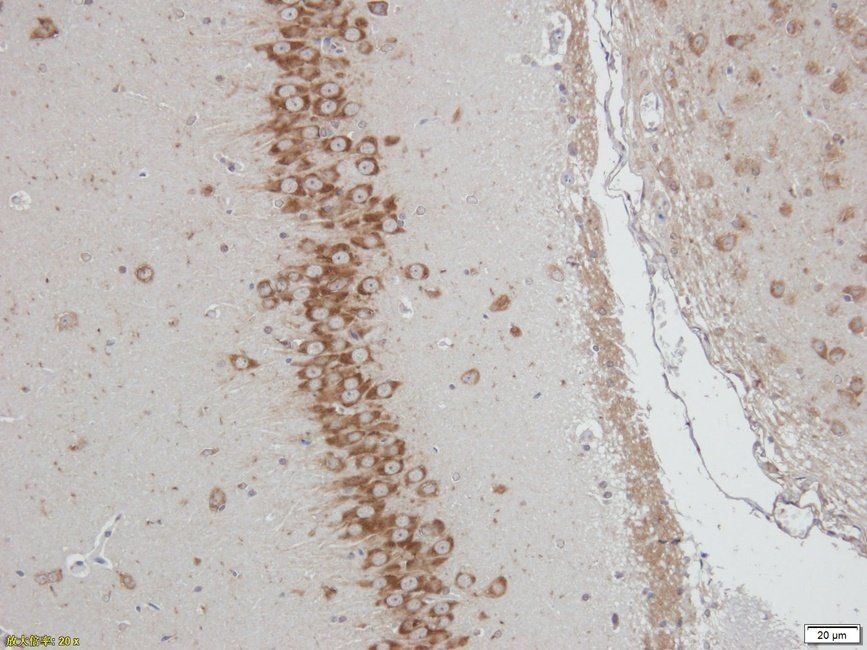
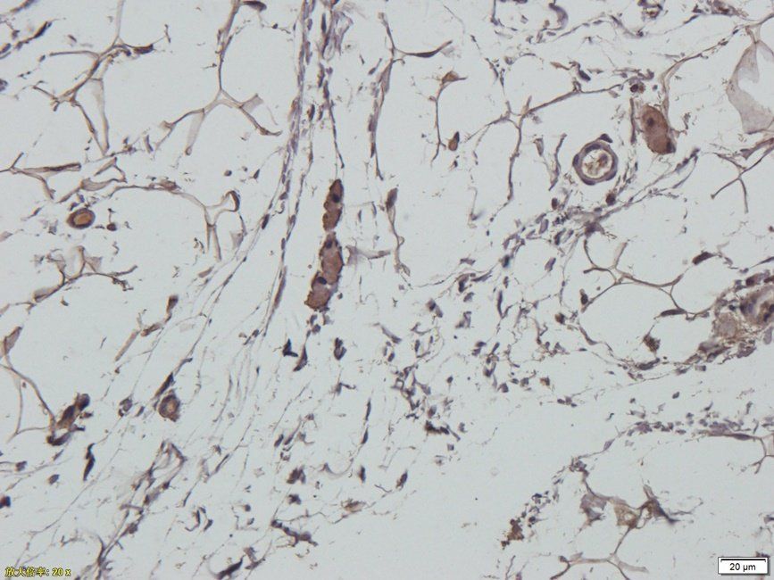
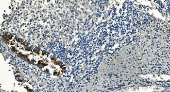
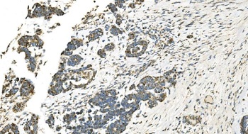
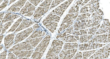
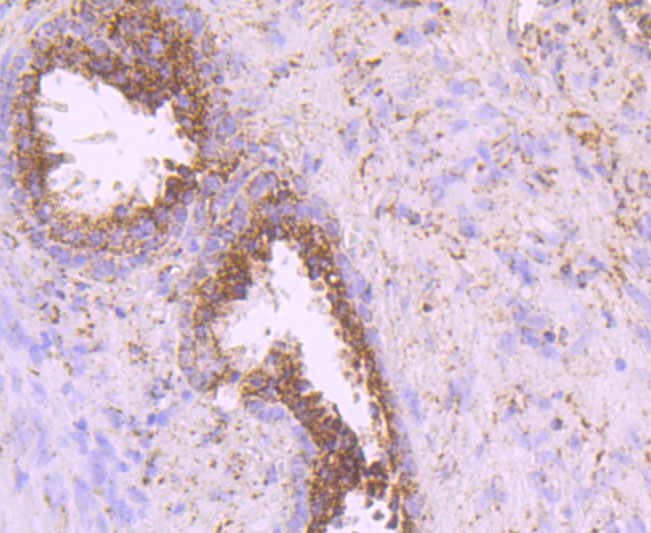
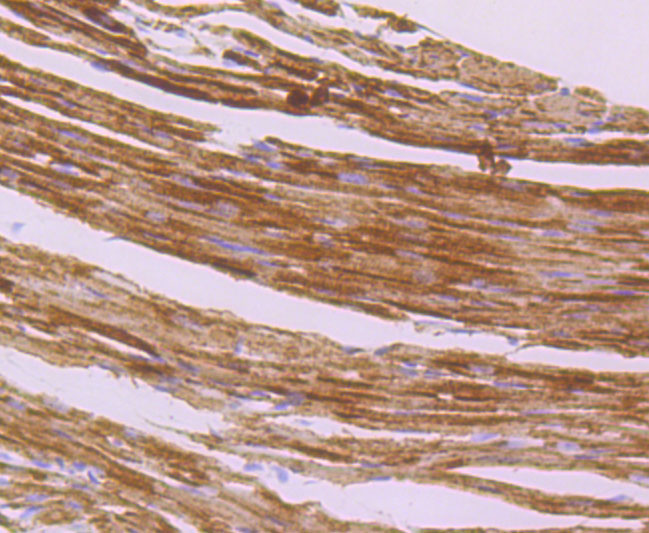
IHC analysis of IDH2 using anti-IDH2 antibody.IDH2 was detected in frozen section of rat small intestine tissue.
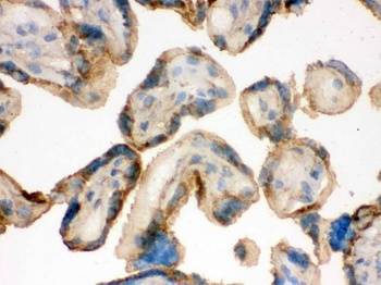
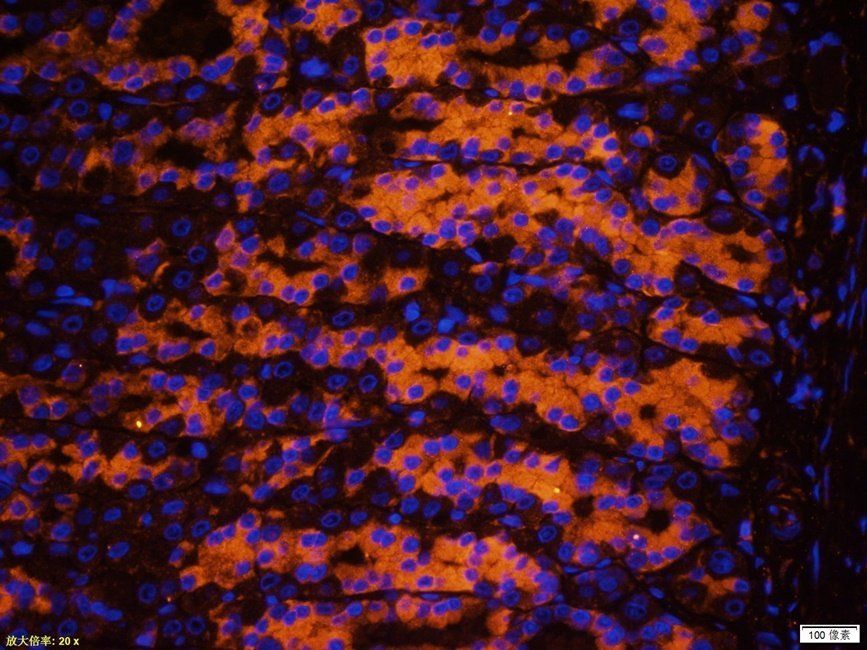
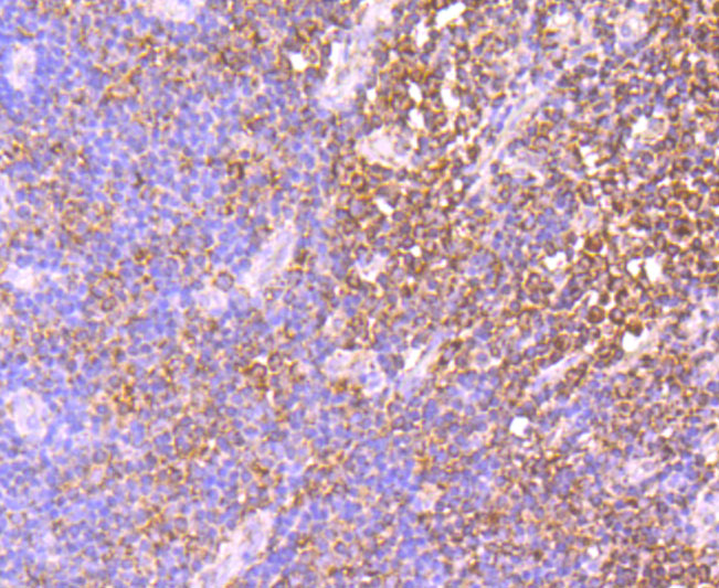
IHC analysis of IDH2 using anti-IDH2 antibody.IDH2 was detected in frozen section of human placenta tissue.
IHC analysis of IDH2 using anti-IDH2 antibody.IDH2 was detected in frozen section of mouse small intestine tissue.
IHC analysis of IDH2 using anti-IDH2 antibody.IDH2 was detected in immunocytochemical section of A549 cell.
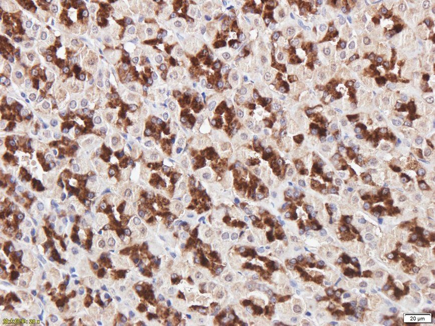
IHC analysis of IDH2 using anti-IDH2 antibody.IDH2 was detected in immunocytochemical section of Hep1-6 cell.
- Item 1 of 9
- Item 1 of 10
IDH2 Antibody (monoclonal, 2H4) [orb692220]
IHC, WB
Human, Mouse, Rat
Mouse
Monoclonal
Unconjugated
10 μg, 100 μg - Item 1 of 8
IDH2 Antibody (monoclonal, 6B13) [orb692221]
FC, ICC, IF, IHC, WB
Human, Mouse, Rat
Mouse
Monoclonal
Unconjugated
10 μg, 100 μg - Item 1 of 7
- Item 1 of 5
IDH2 Antibody [orb1256539]
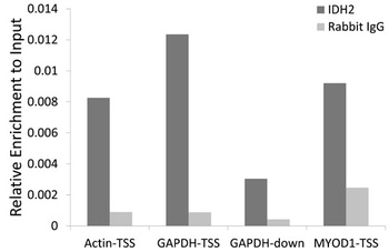
ChIP, IF, IHC, IP, WB
Human, Mouse, Rat
Rabbit
Polyclonal
Unconjugated
100 μl
Submit a review
Filter by Rating

- 5 stars

- 4 stars
- 3 stars
- 2 stars
- 1 stars
