You have no items in your shopping cart.
Cart summary
Item 1 of 3
Item 1 of 3
HSPD1 Antibody (Center)
Catalog Number: orb1788345

| Catalog Number | orb1788345 |
|---|---|
| Category | Antibodies |
| Description | Purified Rabbit Polyclonal Antibody (Pab) |
| Target | This HSPD1 antibody is generated from rabbits immunized with a KLH conjugated synthetic peptide between 187-215 amino acids from the Central region of human HSPD1. |
| Clonality | Polyclonal |
| Species/Host | Rabbit |
| Isotype | Rabbit IgG |
| Conjugation | Unconjugated |
| Reactivity | Human, Mouse |
| Predicted Reactivity | Hamster, Rat |
| Form/Appearance | Purified polyclonal antibody supplied in PBS with 0.09% (W/V) sodium azide. This antibody is prepared by Saturated Ammonium Sulfate (SAS) precipitation followed by dialysis against PBS. |
| Immunogen | 187-215 aa |
| UniProt ID | P10809 |
| MW | 61055 Da |
| Tested applications | IF, WB |
| Dilution range | IF: 1:10~50, IF: 1:100, WB: 1:1000 |
| Antibody Type | Primary Antibody |
| Storage | Maintain refrigerated at 2-8°C for up to 2 weeks. For long term storage store at -20°C in small aliquots to prevent freeze-thaw cycles |
| Alternative names | HSPD1; HSP60; 60 kDa heat shock protein, mitochond Read more... |
| Research Area | Cell Biology, Immunology & Inflammation, Infectiou Read more... |
| Note | For research use only |
| NCBI | NP_002147.2, NP_955472.1 |
| Expiration Date | 12 months from date of receipt. |
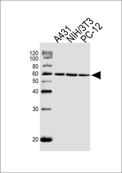
Western blot analysis of lysates from A431, mouse NIH/3T3, rat PC-12 cell line (from left to right), using HSPD1 Antibody (Center). Diluted at 1:1000 at each lane. A goat anti-rabbit IgG H&L (HRP) at 1:10000 dilution was used as the secondary antibody. Lysates at 20ug per lane.
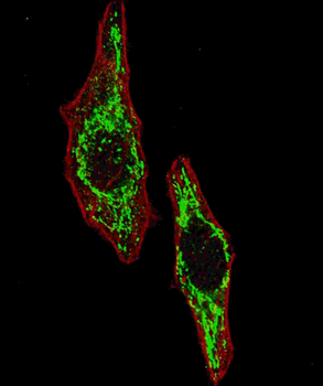
Fluorescent image of U251 cells stained with HSPD1 (Center) antibody. U251 cells were fixed with 4% PFA (20 min), permeabilized with Triton X-100 (0.2%, 30 min). Cells were then incubated with HSPD1 (Center) primary antibody (1: 100, 2 h at room temperature). For secondary antibody, Alexa Fluor 488 conjugated donkey anti-rabbit antibody (green) was used (1:1000, 1h). Cytoplasmic actin was counterstained with Alexa Fluor 555 (red) conjugated Phalloidin (5.25 μM, 25 min).
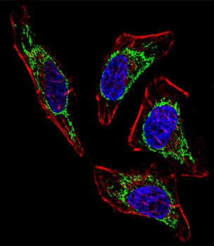
Fluorescent confocal image of Hela cell stained with HSPD1 Antibody (Center). Hela cells were fixed with 4% PFA (20 min), permeabilized with Triton X-100 (0.1%, 10 min), then incubated with HSPD1 primary antibody (1:25, 1 h at 37°C). For secondary antibody, Alexa Fluor 488 conjugated donkey anti-rabbit antibody (green) was used (1:400, 50 min at 37°C).Cytoplasmic actin was counterstained with Alexa Fluor 555 (red) conjugated Phalloidin (7units/ml, 1 h at 37°C). Nuclei were counterstained with DAPI (blue) (10 µg/ml, 10 min). HSPD1 immunoreactivity is localized to Mitochondria significantly.
- Item 1 of 3
HSPD1 Antibody (Center) [orb1931390]
IF, WB
Hamster, Mouse, Rat
Human
Rabbit
Polyclonal
Unconjugated
100 μl, 50 μl


