You have no items in your shopping cart.
Cart summary
Item 1 of 4
Item 1 of 4
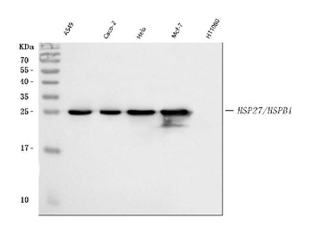
HSPB1 Antibody
Catalog Number: orb1252760
| Catalog Number | orb1252760 |
|---|---|
| Category | Antibodies |
| Description | HSPB1 Antibody |
| Species/Host | Mouse |
| Clonality | Monoclonal |
| Clone Number | G3.1 |
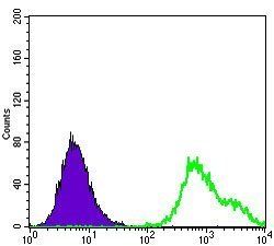
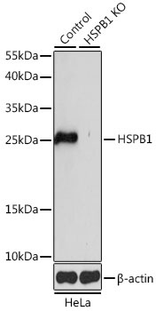
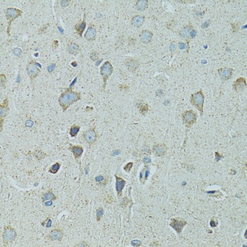
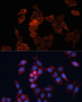
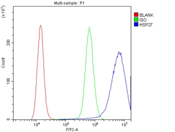
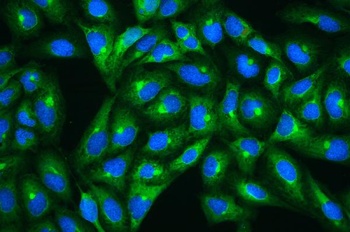
| Tested applications | FC, IF, IHC, WB |
| Reactivity | Gallus, Human, Mouse, Primate, Rat, Sheep |
| Isotype | IgG1, kappa |
| Immunogen | Partially purified human HSP27 (earlier called 24K) from breast cancer MCF-7 cells was used as the immunogen for this antibody. |
| Concentration | 0.2 mg/mL |
| Dilution range | Flow Cytometry: 0.5-1 ug/million cellsIF: 0.5-1 ug/mlWB: 0.25-0.5 ug/mlIHC (FFPE): 0.5-1 ug/ml for 30 minutes at RT (1)Prediluted format : incubate for 30 min at RT (2)The concentration stated for each application is a general starting point. Variations in protocols, secondaries and substrates may require the antibody to be titered up or down for optimal performance.1. Staining of formalin-fixed tissues is enhanced by boiling tissue sections in 10mM Citrate Buffer, pH 6.0, for 10-20 min followed by cooling at RT for 20 minutes.2. The prediluted format is supplied in a dropper bottle and is optimized for use in IHC. After epitope retrieval step (if required), drip mAb solution onto the tissue section and incubate at RT for 30 min. |
| Form/Appearance | Liquid |
| Conjugation | Unconjugated |
| Target | HSPB1 |
| UniProt ID | P04792 |
| Storage | Aliquot and Store at 2-8°C. Avoid freez-thaw cycles. |
| Buffer/Preservatives | PBS with 0.1 mg/ml rAlbumin and 0.05% sodium azide |
| Alternative names | Heat shock protein beta-1, HspB1, 28 kDa heat shoc Read more... |
| Note | For research use only |
| Application notes | Flow Cytometry: 0.5-1 ug/million cellsIF: 0.5-1 ug/mlWB: 0.25-0.5 ug/mlIHC (FFPE): 0.5-1 ug/ml for 30 minutes at RT (1)Prediluted format : incubate for 30 min at RT (2)The concentration stated for each application is a general starting point. Variations in protocols, secondaries and substrates may require the antibody to be titered up or down for optimal performance.1. Staining of formalin-fixed tissues is enhanced by boiling tissue sections in 10mM Citrate Buffer, pH 6.0, for 10-20 min followed by cooling at RT for 20 minutes.2. The prediluted format is supplied in a dropper bottle and is optimized for use in IHC. After epitope retrieval step (if required), drip mAb solution onto the tissue section and incubate at RT for 30 min. |
| Expiration Date | 12 months from date of receipt. |
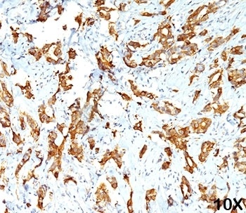
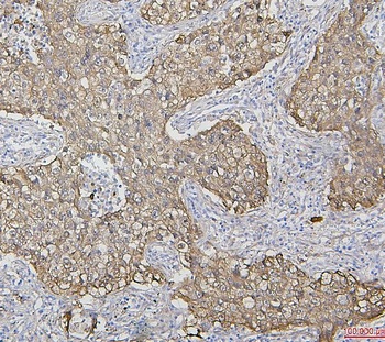
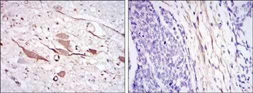
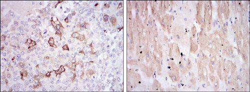
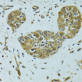
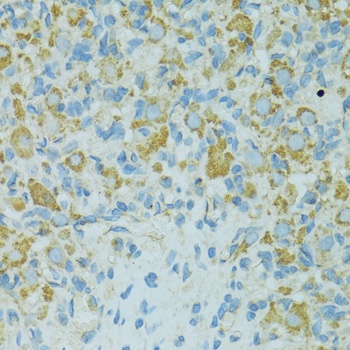
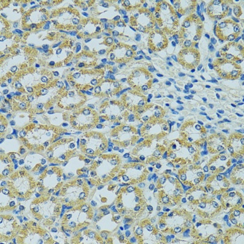
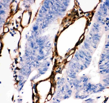
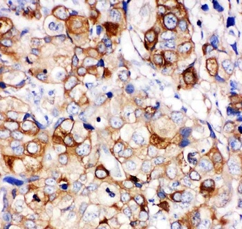
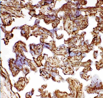
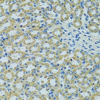
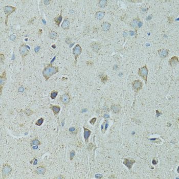
IHC testing of human breast carcinoma (10X) stained with HSP27 antibody (G3.1).
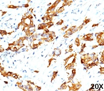
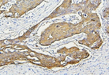
IHC testing of breast carcinoma (20X) stained with HSP27 antibody (G3.1).
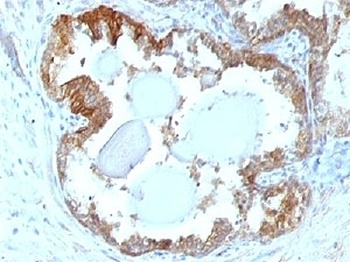
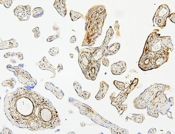
IHC testing of FFPE human prostate carcinoma and HSP27 antibody.
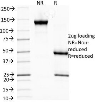
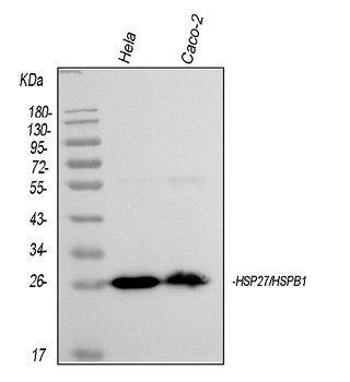
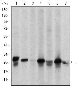
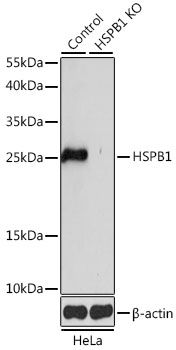
SDS-PAGE Analysis of Purified, BSA-Free HSP27 Antibody (clone G3.1). Confirmation of Integrity and Purity of the Antibody.
- Item 1 of 7
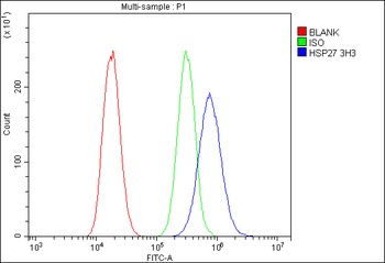
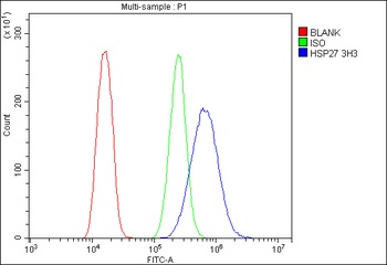
Hsp27/HSPB1 Antibody (monoclonal, 3H3) [orb570310]
FC, ICC, IF, IHC, WB
Human
Mouse
Monoclonal
Unconjugated
10 μg, 100 μg - Item 1 of 6
- Item 1 of 6
- Item 1 of 6
Hsp27/HSPB1 Antibody [orb215991]
FC, ICC, IF, IHC, IHC-Fr, WB
Human
Rabbit
Polyclonal
Unconjugated
10 μg, 100 μg - Item 1 of 5


Submit a review
Filter by Rating

- 5 stars

- 4 stars
- 3 stars
- 2 stars
- 1 stars