You have no items in your shopping cart.
Cart summary
Item 1 of 4
Item 1 of 4
HSPA8 Antibody
Catalog Number: orb1264542
| Catalog Number | orb1264542 |
|---|---|
| Category | Antibodies |
| Description | HSPA8 Antibody |
| Species/Host | Rabbit |
| Clonality | Polyclonal |
| Tested applications | FC, IF, IHC-P, WB |
| Predicted Reactivity | Bovine, Gallus, Hamster, Mouse, Rat |
| Reactivity | Human |
| Isotype | Rabbit Ig |
| Immunogen | This HSPA8 antibody is generated from rabbits immunized with a KLH conjugated synthetic peptide between 82-110 amino acids from the N-terminal region of human HSPA8. |
| Concentration | batch dependent |
| Dilution range | For WB starting dilution is: 1:1000For IHC-P starting dilution is: 1:10~50For IF starting dilution is: 1:10~50For FACS starting dilution is: 1:10~50 |
| Form/Appearance | Liquid |
| Conjugation | Unconjugated |
| MW | 71 kDa |
| Target | HSPA8 |
| UniProt ID | P11142 |
| NCBI | P11142 |
| Storage | Store at 4°C for three months and -20°C, stable for up to one year. As with all antibodies care should be taken to avoid repeated freeze thaw cycles. Antibodies should not be exposed to prolonged high temperatures. |
| Buffer/Preservatives | Supplied in PBS with 0.09% (W/V) sodium azide. |
| Alternative names | Heat shock cognate 71 kDa protein, Heat shock 70 k Read more... |
| Note | For research use only |
| Application notes | For WB starting dilution is: 1:1000For IHC-P starting dilution is: 1:10~50For IF starting dilution is: 1:10~50For FACS starting dilution is: 1:10~50 |
| Expiration Date | 12 months from date of receipt. |
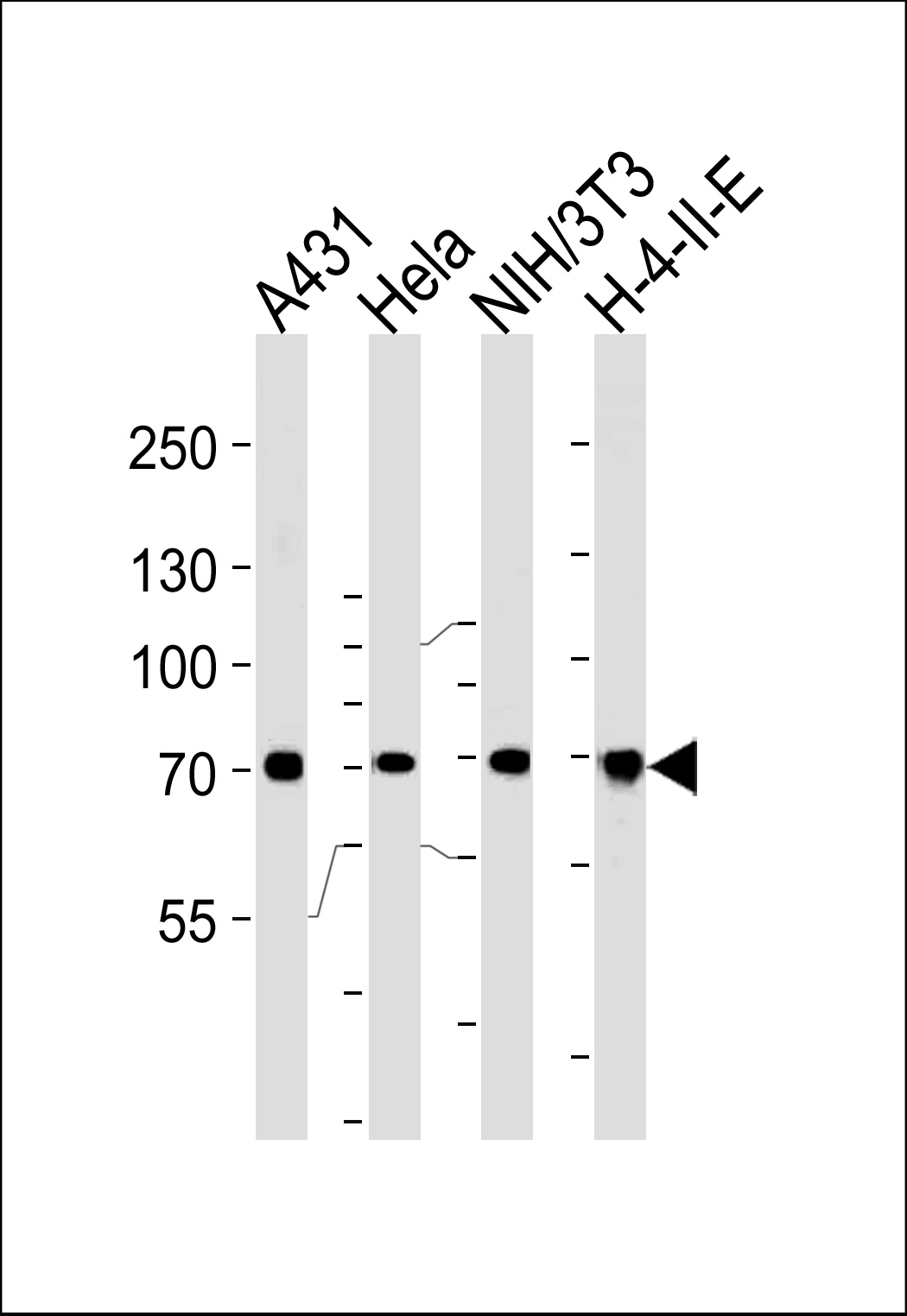
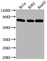
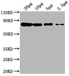
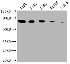
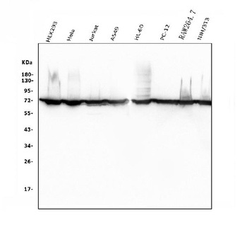
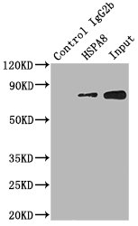
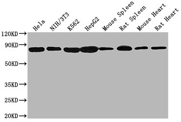
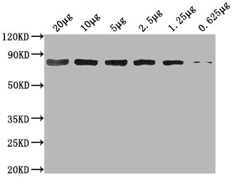
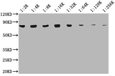
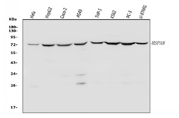
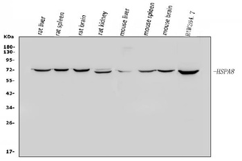
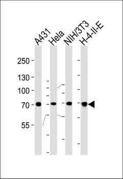
Western blot analysis of lysates from A431, Hela, mouse NIH/3T3, H-4-II-E cell line (from left to right), using HSPA8 Antibody.AP2872a was diluted at 1:1000 at each lane.
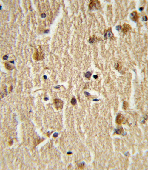
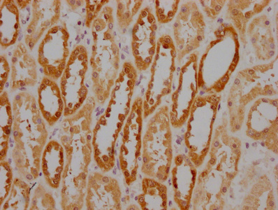
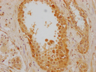
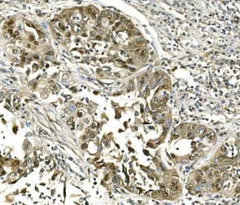
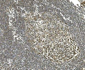
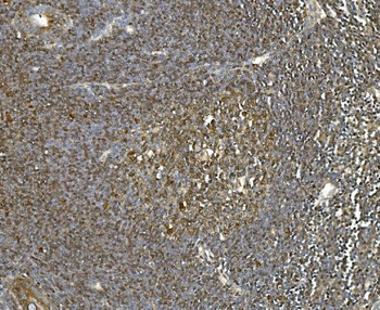
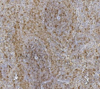
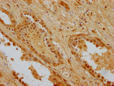
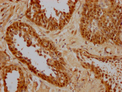
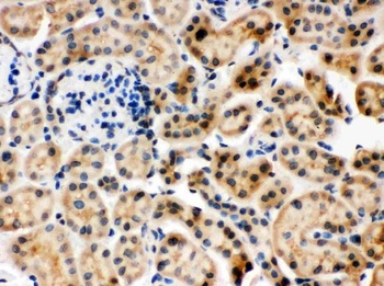
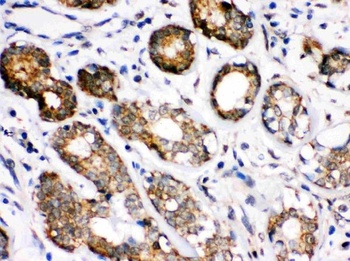
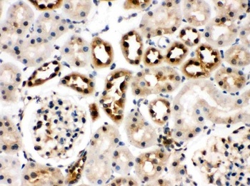
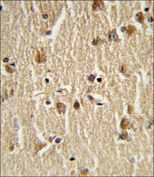
Formalin-fixed and paraffin-embedded human brain tissue reacted with HSPA8 Antibody (N-term), which was peroxidase-conjugated to the secondary antibody, followed by DAB staining.
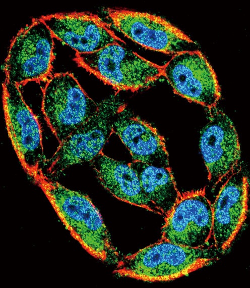
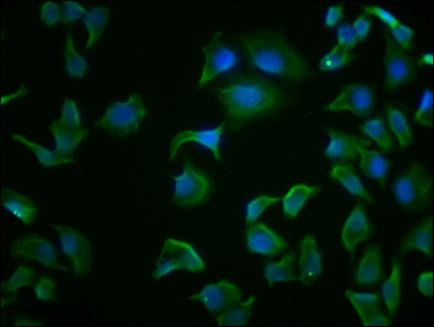
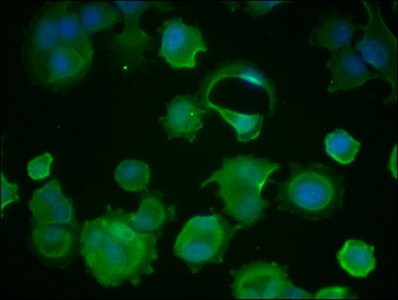
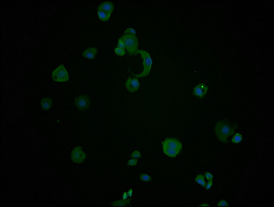
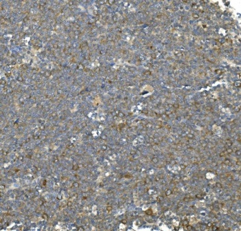
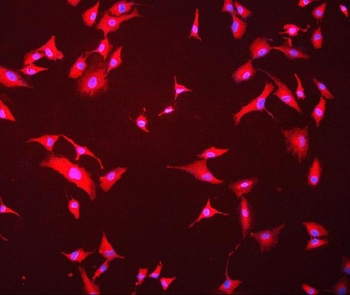
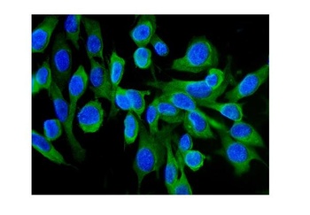
Confocal immunofluorescent analysis of HSPA8 Antibody with A2058 cell followed by Alexa Fluor 488-conjugated goat anti-rabbit lgG (green). Actin filaments have been labeled with Alexa Fluor 555 phalloidin (red).DAPI was used to stain the cell nuclear (blue).
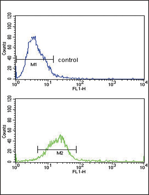
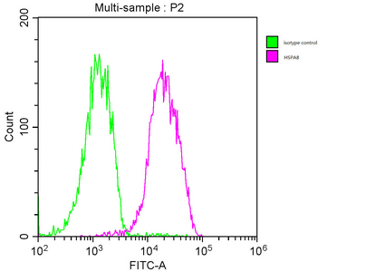
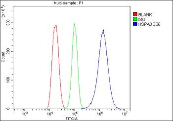
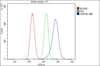
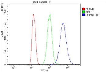
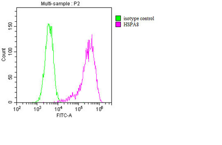
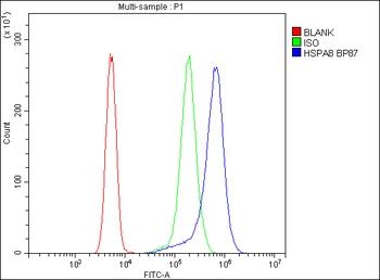
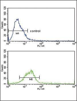
Flow cytometric analysis of Hela cells (bottom histogram) compared to a negative control cell (top histogram). FITC-conjugated goat-anti-rabbit secondary antibodies were used for the analysis.
- Item 1 of 9
- Item 1 of 10
Hsc70 Antibody(monoclonal, 3B6) [orb623830]
FC, ICC, IF, IHC, WB
Human, Mouse, Rat
Mouse
Monoclonal
Unconjugated
10 μg, 100 μg - Item 1 of 7
HSPA8 antibody [orb688870]
ELISA, FC, IHC, IP, WB
Human, Mouse, Rat
Mouse
Monoclonal
Unconjugated
50 μl, 100 μl - Item 1 of 7
Hsc70/HSPA8 Antibody [orb315149]
FC, ICC, IF, IHC, WB
Human, Mouse, Rat
Rabbit
Polyclonal
Unconjugated
10 μg, 100 μg - Item 1 of 4
HSPA8 antibody [orb34255]
FC, IF, IHC-P, WB
Equine, Hamster
Human, Mouse, Rat
Rabbit
Polyclonal
Unconjugated
80 μl

Submit a review
Filter by Rating

- 5 stars

- 4 stars
- 3 stars
- 2 stars
- 1 stars