You have no items in your shopping cart.
Cart summary
Item 1 of 6
Item 1 of 6
HLA-DRB1 Antibody (MHC II)
Catalog Number: orb2638570

| Catalog Number | orb2638570 |
|---|---|
| Category | Antibodies |
| Description | HLA-DRB1 binds peptides derived from antigens that access the endocytic route of antigen presenting cells (APC) and presents them on the cell surface for recognition by the CD4 T-cells. [UniProt] |
| Clonality | Monoclonal |
| Species/Host | Mouse |
| Isotype | Mouse IgG2b, kappa |
| Conjugation | Unconjugated |
| Reactivity | Human |
| Buffer/Preservatives | 0.2 mg/ml in 1X PBS with 0.1 mg/ml rAlbumin (US sourced) and 0.05% sodium azide |
| Purity | Protein G affinity chromatography |
| Immunogen | Activated human peripheral blood mononuclear cells were used as the immunogen for the HLA-DRB1 antibody. |
| UniProt ID | P01911 |
| Tested applications | FACS, IF, IHC-P, WB |
| Dilution range | Flow cytometry: 1-2ug/million cells,Immunofluorescence: 1-3ug/ml,Western blot: 2-4ug/ml,Immunohistochemistry (FFPE): 1-2ug/ml for 30 min at RT |
| Application notes | Optimal dilution of the HLA-DRB1 antibody should be determined by the researcher.1. Staining of formalin-fixed tissues requires boiling tissue sections in 10mM Citrate buffer, pH 6.0, for 10-20 min followed by cooling at RT for 20 min2. The prediluted format is supplied in a dropper bottle and is optimized for use in IHC. After epitope retrieval step (if required), drip mAb solution onto the tissue section and incubate at RT for 30 min. |
| Antibody Type | Primary Antibody |
| Clone Number | LN3 |
| Formula | 0.2 mg/ml in 1X PBS with 0.1 mg/ml BSA (US sourced) and 0.05% sodium azide |
| Storage | Maintain refrigerated at 2-8°C for up to 2 weeks. For long term storage store at -20°C in small aliquots to prevent freeze-thaw cycles. |
| Hazard Information | This HLA-DRB1 antibody is available for research use only. |
| Note | For research use only |
| Expiration Date | 12 months from date of receipt. |

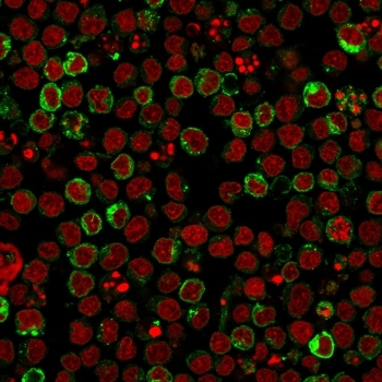
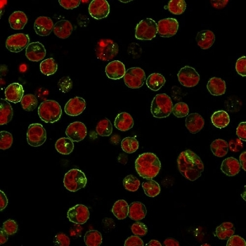
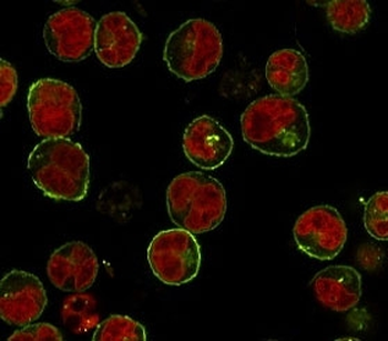
Immunofluorescent staining of human Ramos cells with HLA-DRB1 antibody (clone LN3, green) and Reddot nuclear stain (red).

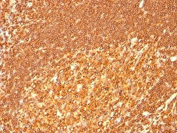
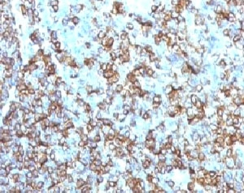
IHC staining of FFPE human tonsil tissue with HLA-DRB1 antibody (clone LN3). HIER: boil tissue sections in pH9 10mM Tris with 1mM EDTA for 20 min and allow to cool before testing.
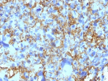
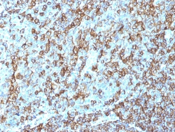
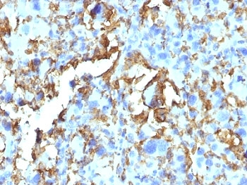
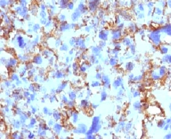
IHC staining of FFPE human histiocytoma tissue with HLA-DRB1 antibody (clone LN3). HIER: boil tissue sections in pH9 10mM Tris with 1mM EDTA for 20 min and allow to cool before testing.

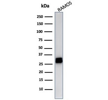
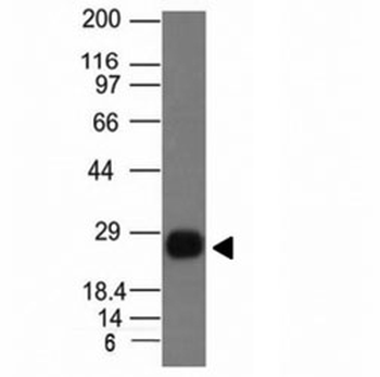
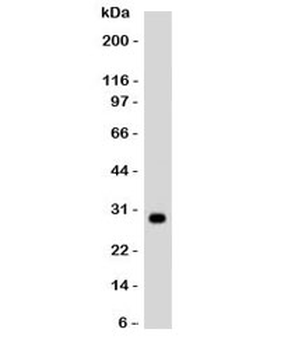
Western blot testing of human Ramos cell lysate with HLA-DRB1 antibody (clone LN3). Expected molecular weight ~30 kDa.
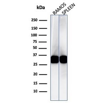
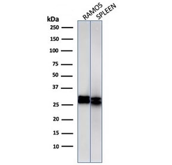
Western blot testing of human Ramos and spleen cell lysate with HLA-DRB1 antibody (clone LN3). Expected molecular weight ~30 kDa.
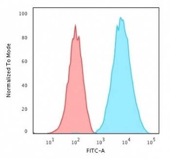
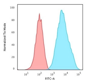
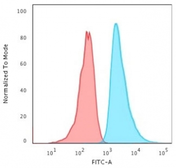
Flow cytometry staining of human Raji cells with HLA-DRB1 antibody; Red = isotype control, Blue = HLA-DRB1 antibody.
- Item 1 of 6
- Item 1 of 6
- Item 1 of 6
- Item 1 of 5
- Item 1 of 5