You have no items in your shopping cart.
Cart summary
Item 1 of 6
Item 1 of 6
GLG1 Antibody (C-term)
Catalog Number: orb1927695

| Catalog Number | orb1927695 |
|---|---|
| Category | Antibodies |
| Description | Affinity Purified Rabbit Polyclonal Antibody (Pab) |
| Target | This GLG1 antibody is generated from rabbits immunized with a KLH conjugated synthetic peptide between 1152-1179 amino acids from the C-terminal region of human GLG1. |
| Clonality | Polyclonal |
| Species/Host | Rabbit |
| Isotype | Rabbit IgG |
| Conjugation | Unconjugated |
| Reactivity | Human, Mouse |
| Predicted Reactivity | Hamster, Rat |
| Form/Appearance | Purified polyclonal antibody supplied in PBS with 0.09% (W/V) sodium azide. This antibody is purified through a protein A column, followed by peptide affinity purification. |
| UniProt ID | Q92896 |
| MW | 134552 Da |
| Tested applications | IHC-P, WB |
| Dilution range | WB: 1:2000, WB: 1:1000, WB: 1:1000, WB: 1:2000, WB: 1:2000, IHC-P: 1:50~100 |
| Clone Number | RB24297 |
| Storage | Maintain refrigerated at 2-8°C for up to 2 weeks. For long term storage store at -20°C in small aliquots to prevent freeze-thaw cycles |
| Alternative names | Golgi apparatus protein 1, CFR-1, Cysteine-rich fi Read more... |
| Note | For research use only |
| NCBI | NP_001139138.1, NP_036333.2, NP_001139139.1 |
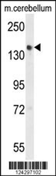
Western blot analysis of GLG1 Antibody (C-term) in mouse cerebellum tissue lysates (35 ug/lane). GLG1 (arrow) was detected using the purified Pab.
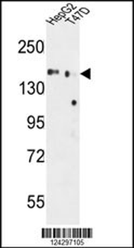
GLG1 Antibody (C-term) western blot analysis in HepG2, T47D cell line lysates (35 ug/lane).This demonstrates the GLG1 antibody detected the GLG1 protein (arrow).
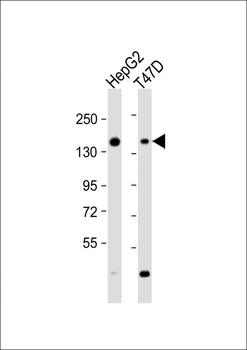
All lanes: Anti-GLG1 Antibody (C-term) at 1:2000 dilution. Lane 1: HepG2 whole cell lysate. Lane 2: T47D whole cell lysate. Lysates/proteins at 20 µg per lane. Secondary Goat Anti-Rabbit IgG, (H+L), Peroxidase conjugated at 1/10000 dilution. Predicted band size: 136 kDa. Blocking/Dilution buffer: 5% NFDM/TBST.

GLG1 Antibody (C-term) IHC analysis in formalin fixed and paraffin embedded skeletal muscle followed by peroxidase conjugation of the secondary antibody and DAB staining. This data demonstrates the use of the GLG1 Antibody (C-term) for immunohistochemistry. Clinical relevance has not been evaluated.
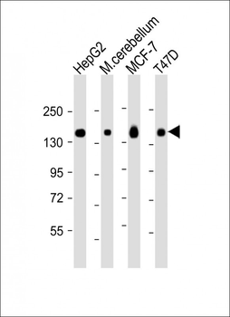
All lanes: Anti-GLG1 Antibody (C-term) at 1:2000 dilution. Lane 1: HepG2 whole cell lysate. Lane 2: mouse cerebellum lysate. Lane 3: MCF-7 whole cell lysate. Lane 4: T47D whole cell lysate. Lysates/proteins at 20 µg per lane. Secondary Goat Anti-Rabbit IgG, (H+L), Peroxidase conjugated at 1/10000 dilution. Predicted band size: 136 kDa. Blocking/Dilution buffer: 5% NFDM/TBST.
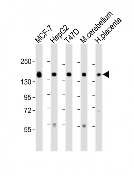
All lanes: Anti-GLG1 Antibody (C-term) at 1:2000 dilution. Lane 1: MCF-7 lysate. Lane 2: HepG2 whole cell lysate. Lane 3: T47D whole cell lysate. Lane 4: mouse cerebellum lysate. Lane 5: human placenta lysate. Lysates/proteins at 20 µg per lane. Secondary Goat Anti-Rabbit IgG, (H+L), Peroxidase conjugated at 1/10000 dilution. Predicted band size: 136 kDa. Blocking/Dilution buffer: 5% NFDM/TBST.
