You have no items in your shopping cart.
Cart summary
Item 1 of 4
Item 1 of 4
GFAP Antibody / Glial Fibrillary Acidic Protein
Catalog Number: orb1825069

| Catalog Number | orb1825069 |
|---|---|
| Category | Antibodies |
| Description | GFAP is specifically found in astroglia. GFAP is a very popular marker for localizing benign astrocyte and neoplastic cells of glial origin in the central nervous system. Antibody to GFAP is useful in differentiating primary gliomas from metastatic lesions in the brain and for documenting astrocytic differentiation in tumors outside the CNS. |
| Species/Host | Mouse |
| Clonality | Monoclonal |
| Clone Number | GFAP/6878 |
| Tested applications | IHC-P, WB |
| Reactivity | Human |
| Isotype | Mouse IgG1, kappa |
| Immunogen | A recombinant partial protein sequence (within amino acids 150-250) from the human protein was used as the immunogen for the GFAP antibody. |
| Antibody Type | Primary Antibody |
| Dilution range | Immunohistochemistry (FFPE): 1-2ug/ml for 30 min at RT,Western blot: 1-2ug/ml |
| Conjugation | Unconjugated |
| Formula | 0.2 mg/ml in 1X PBS with 0.1 mg/ml BSA (US sourced), 0.05% sodium azide |
| Hazard Information | This GFAP antibody is available for research use only. |
| UniProt ID | P14136 |
| Storage | Aliquot the GFAP antibody and store frozen at -20°C or colder. Avoid repeated freeze-thaw cycles. |
| Note | For research use only |
| Expiration Date | 12 months from date of receipt. |

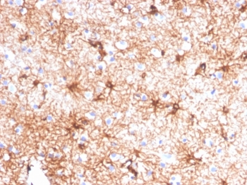
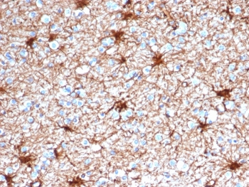
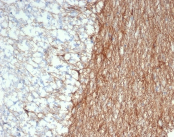
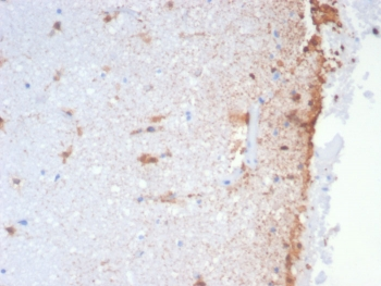
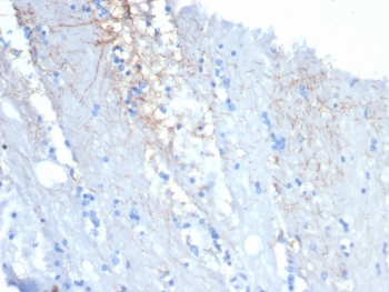
IHC staining of FFPE human cerebellum tissue with GFAP antibody (clone GFAP/6878). HIER: boil tissue sections in pH9 10 mM Tris with 1 mM EDTA for 20 min and allow to cool before testing.
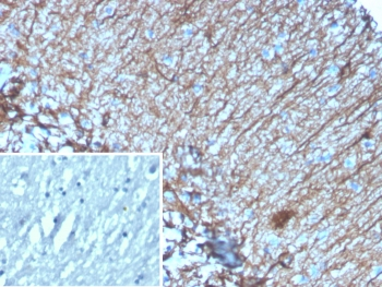
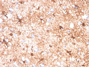
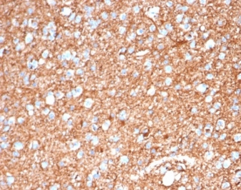
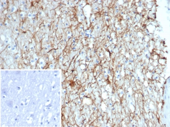
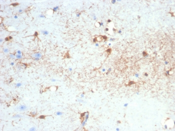
IHC staining of FFPE human brain tissue with GFAP antibody (clone GFAP/6878). Inset: PBS used in place of primary Ab (secondary Ab negative control). HIER: boil tissue sections in pH9 10 mM Tris with 1 mM EDTA for 20 min and allow to cool before testing.
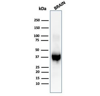
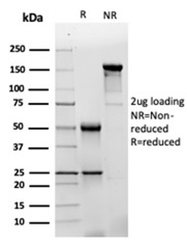
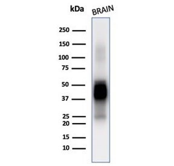
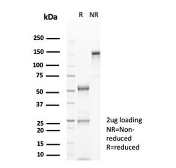
Western blot testing of human brain tissue lysate with GFAP antibody (clone GFAP/6878).
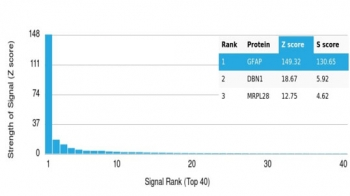
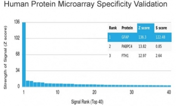
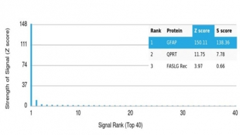
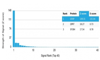
Analysis of a HuProt (TM) microarray containing more than 19000 full-length human proteins using GFAP antibody (clone GFAP/6878). Z- and S- Score: The Z-score represents the strength of a signal that a monoclonal antibody (in combination with a fluorescently-tagged anti-IgG secondary antibody) produces when binding to a particular protein on the HuProt (TM) array. Z-scores are described in units of standard deviations (SD's) above the mean value of all signals generated on that array. If targets on HuProt (TM) are arranged in descending order of the Z-score, the S-score is the difference (also in units of SD's) between the Z-score. S-score therefore represents the relative target specificity of a mAb to its intended target. A mAb is considered to specific to its intended target, if the mAb has an S-score of at least 2.5. For example, if a mAb binds to protein X with a Z-score of 43 and to protein Y with a Z-score of 14, then the S-score for the binding of that mAb to protein X is equal to 29.
- Item 1 of 5
GFAP Antibody / Glial Fibrillary Acidic Protein [orb1151443]
IHC-P
Human
Mouse
Monoclonal
Unconjugated
100 μg - Item 1 of 5
GFAP Antibody / Glial Fibrillary Acidic Protein [orb1825010]
IHC-P, WB
Human
Mouse
Monoclonal
Unconjugated
100 μg - Item 1 of 5
GFAP Antibody / Glial Fibrillary Acidic Protein [orb1825011]
IHC-P, WB
Human
Mouse
Monoclonal
Unconjugated
20 μg - Item 1 of 5
GFAP Antibody / Glial Fibrillary Acidic Protein [orb1825012]
IHC-P, WB
Human
Mouse
Monoclonal
Unconjugated
100 μg - Item 1 of 5
GFAP Antibody / Glial Fibrillary Acidic Protein [orb1825064]
IHC-P
Bovine, Canine, Equine, Human
Mouse
Monoclonal
Unconjugated
100 μg