You have no items in your shopping cart.
Cart summary
Item 1 of 3
Item 1 of 3
FAS Antibody
Catalog Number: orb1671856

| Catalog Number | orb1671856 |
|---|---|
| Category | Antibodies |
| Description | FAS Antibody |
| Clonality | Recombinant |
| Clone Number | R-125224 |
| Tested applications | Activation, ELISA, FC |
| Reactivity | Human |
| Isotype | IgG kappa |
| Immunogen | R-125224 is generated by the humanization of the murine HFE7A anti-Fas antibody by grafting the CDR regions to the framework regions of the human 8E10 antibody and substituting key framework residues from the murine antibody into the 8E10 sequence. The original HFE7A was derived from a hybridoma cell line generated by the fusion of NS1 myeloma cells with splenocytes from Fas-deficient mice which had been immunized with partially purified recombinant human Fas-AIC2A chimera protein consisting of the extracellular region of human Fas antigen (aa -16 to 150) and the extracellular region of the murine IL-3 receptor AIC2 (aa 3-423). The HFE7A hybridoma was selected after screening by flow cytometry for the production of antibodies with the ability to bind to the WR19L12a transformed murine T cell lymphoma cell line expressing human Fas or the L5178YA1 cell line expressing murine Fas, but not to the parental WR19L or L5178Y cells. |
| Concentration | batch dependent |
| Conjugation | Unconjugated |
| Target | FAS |
| UniProt ID | P25445 |
| Storage | Store at 4°C for up to 3 months. For longer storage, aliquot and store at -20°C. |
| Buffer/Preservatives | PBS with 0.02% Proclin 300. |
| Alternative names | CD95, h-HFE7A, Tumor necrosis factor receptor supe Read more... |
| Note | For research use only |
| Expiration Date | 12 months from date of receipt. |
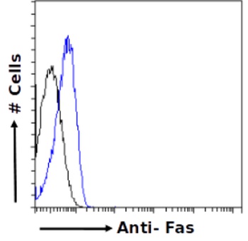
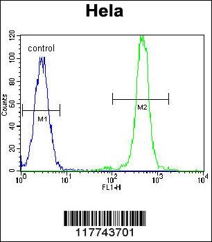
Flow-cytometry using the anti-Fas antibody R-125224. Jurkat cells were fixed using 2% PFA- permeusing 0.5% Triton and stained with unimmunized r antibody (MOPC-21; isotype control - black line) or the r-chimeric version of R-125224 (orb1671856 - blue line) at a dilution of 1:100 for 1h at RT. After washing- bound antibody was detected using a goat anti-r AlexaFluor® 488 antibody at a dilution of 1:1000 and cells analyzed using a FACSCanto flow-cytometer.
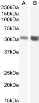
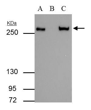
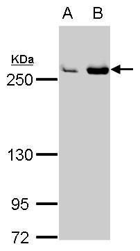
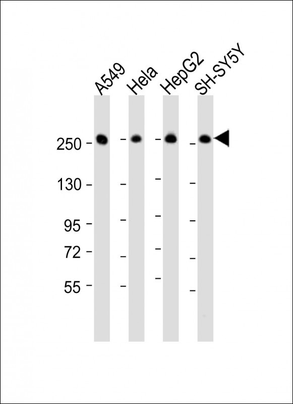
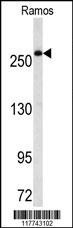
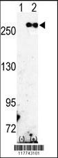
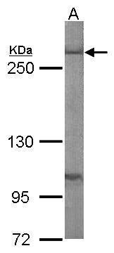
Western Blot using anti-Fas antibody R-125224. Human testis (A) and human ovary (B) lysate samples (35 ug protein in RIPA buffer) were resolved on a 10% SDS PAGE gel and blots probed with the chimeric r version of R-125224 (orb1671856) at 2 ug/ml before detection using an anti-rondary antibody. A primary incubation of 1h was used and protein was detected by chemiluminescence. The expected running size for unmodified Fas is 37.7kDa- but this protein is glycosylated at several positions leading to the observed running size.
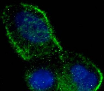
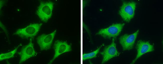
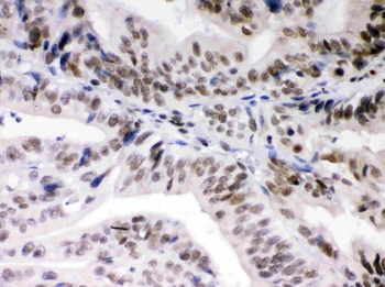
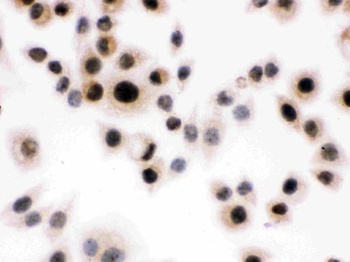
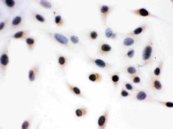
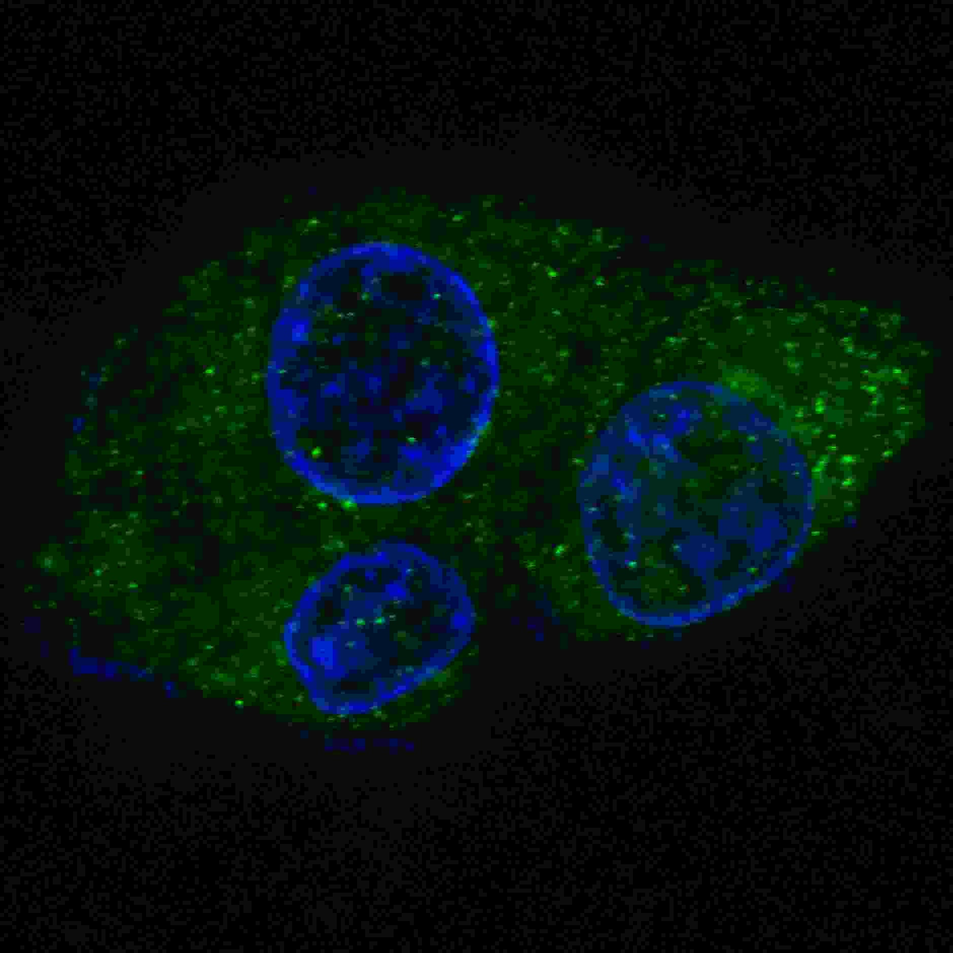
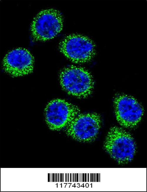
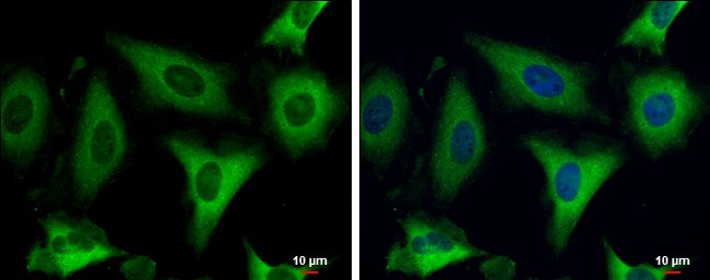
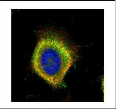
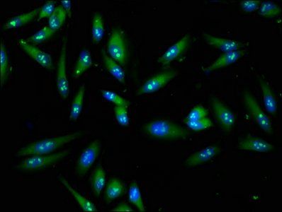
Immunofluorescence staining of fixed MCF7 cells with anti-Fas antibody R-125224. Immunofluorescence analysis of paraformaldehyde fixed MCF7 cells permewith 0.15% Triton and stained with the chimeric mouse IgG1 version of R-125224 (orb1671856) at 10 ug/ml for 1h followed by Alexa Fluor® 488 secondary antibody (2 ug/ml)- showing membrane staining. The nuclear stain is DAPI (blue). Panels show from left-right- top-bottom orb1671856- DAPI- merged channels and an isotype control. The isotype control was stained with an anti-unknown specificity antibody (0) followed by Alexa Fluor® 488 secondary antibody.
- Item 1 of 8
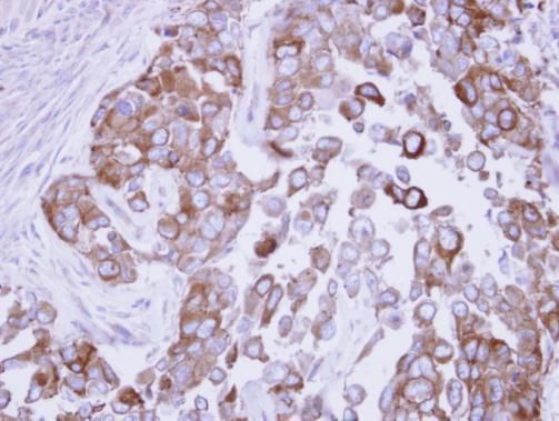
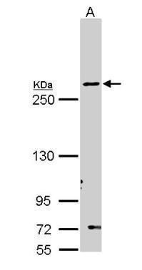
Fatty Acid Synthase antibody [orb556642]
ICC, IHC-Fr, IHC-P, IP, WB
Human, Mouse, Rat
Rabbit
Polyclonal
Unconjugated
100 μl - Item 1 of 8
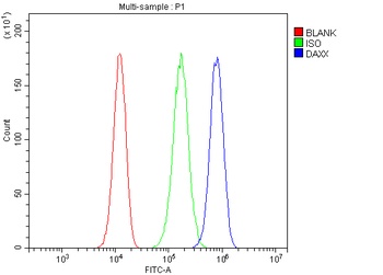
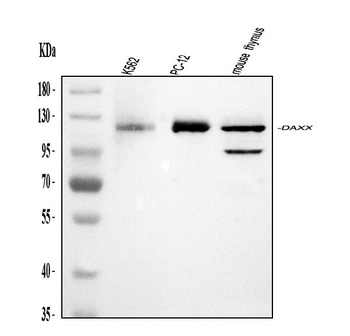
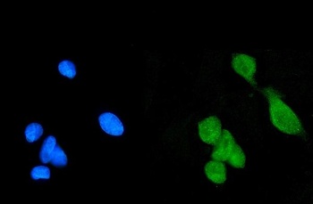
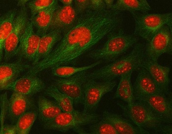
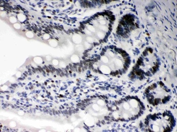
Daxx Antibody [orb308845]
FC, ICC, IF, IHC, WB
Human, Mouse, Rat
Rabbit
Polyclonal
Unconjugated
10 μg, 100 μg - Item 1 of 7
- Item 1 of 7
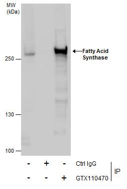
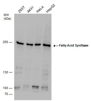
Fatty Acid Synthase antibody [orb556692]
ICC, IHC-P, IP, WB
Human, Mouse
Rabbit
Polyclonal
Unconjugated
100 μl - Item 1 of 5
Submit a review
Filter by Rating

- 5 stars

- 4 stars
- 3 stars
- 2 stars
- 1 stars
