You have no items in your shopping cart.
Cart summary
Item 1 of 5
Item 1 of 5
EPHB1 Antibody
Catalog Number: orb1263085
| Catalog Number | orb1263085 |
|---|---|
| Category | Antibodies |
| Description | EPHB1 Antibody |
| Species/Host | Rabbit |
| Clonality | Polyclonal |
| Tested applications | IHC-P, WB |
| Reactivity | Human, Mouse |
| Isotype | Rabbit Ig |
| Immunogen | This EphB1 antibody is generated from rabbits immunized with a KLH conjugated synthetic peptide between 955-984 amino acids from the C-terminal region of human EphB1. |
| Concentration | batch dependent |
| Dilution range | For WB starting dilution is: 1:1000For IHC-P starting dilution is: 1:50~100 |
| Form/Appearance | Liquid |
| Conjugation | Unconjugated |
| MW | 110 kDa |
| Target | EPHB1 |
| UniProt ID | P54762 |
| NCBI | P54762 |
| Storage | Store at 4°C for three months and -20°C, stable for up to one year. As with all antibodies care should be taken to avoid repeated freeze thaw cycles. Antibodies should not be exposed to prolonged high temperatures. |
| Buffer/Preservatives | Supplied in PBS with 0.09% (W/V) sodium azide. |
| Alternative names | Ephrin type-B receptor 1, ELK, EPH tyrosine kinase Read more... |
| Note | For research use only |
| Application notes | For WB starting dilution is: 1:1000For IHC-P starting dilution is: 1:50~100 |
| Expiration Date | 12 months from date of receipt. |

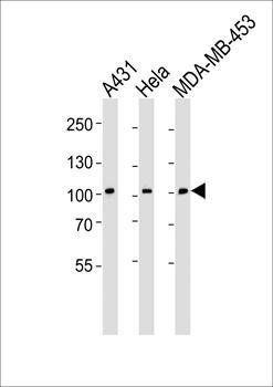
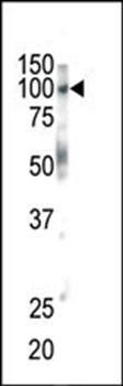
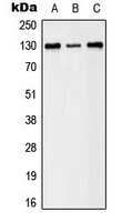
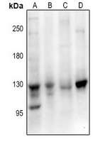
Western blot analysis of lysates from A431, Hela, MDA-MB-453 cell line (from left to right), using EPHB1 Antibody (H970) at 1:1000 at each lane.
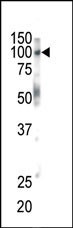
Western blot analysis of anti-EphB1 Pab in mouse brain tissue
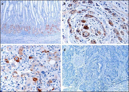
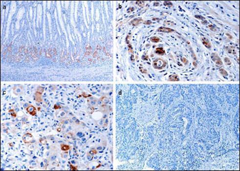
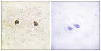
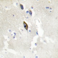
Immunohistochemical analysis of EphB1 in gastric cancer tissues. a EphB1 protein expressed in normal mucosa at the glandular compartment and in a decreasing gradient from the glandular compartment to the foveolar compartment. b EphB1 protein focally positively stained in well-differentiated gastric cancer cells. c EphB1 protein is focally positive in poorly differentiated gastric cancer cells. d Loss of EphB1 expression in gastric cancer cells. (Provided by Jian-dong Wang, Department of Pathology Nanjing Jinling Hospital/Nanjing University School of Medicine)
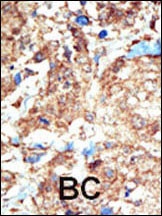
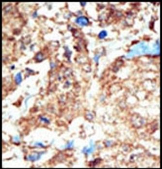
Formalin-fixed and paraffin-embedded human cancer tissue reacted with the primary antibody, which was peroxidase-conjugated to the secondary antibody, followed by DAB staining. BC = breast carcinoma; HC = hepatocarcinoma.
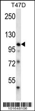
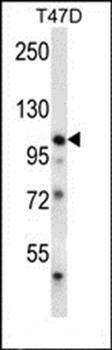
Western blot analysis in T47D cell line lysates (35 ug/lane).
- Item 1 of 5
- Item 1 of 4
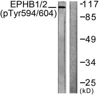
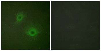
EphB1/2 (phospho-Tyr594/604) antibody [orb764324]
ELISA, IF, WB
Human, Mouse, Rat
Rabbit
Polyclonal
Unconjugated
50ul, 100ul - Item 1 of 4
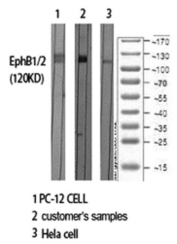
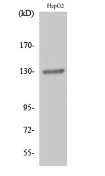
EphB1/2 antibody [orb765148]
ELISA, IF, IHC-P, WB
Human, Mouse, Rat
Rabbit
Polyclonal
Unconjugated
50ul, 100ul - Item 1 of 3
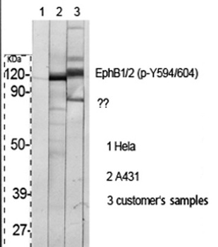
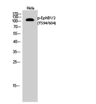
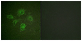
EPHB1/2 (phospho-Y594/604) antibody [orb224166]
IF, WB
Human, Mouse, Rat
Rabbit
Polyclonal
Unconjugated
200 μl, 100 μl, 30 μl - Item 1 of 3
EPHB1/2/3 antibody [orb665861]
IF, IH, WB
Human, Mouse, Rat, Zebrafish
Rabbit
Polyclonal
Unconjugated
200 μl, 100 μl, 30 μl
Submit a review
Filter by Rating

- 5 stars

- 4 stars
- 3 stars
- 2 stars
- 1 stars