You have no items in your shopping cart.
Cart summary
Item 1 of 6
Item 1 of 6
EphA2 antibody
Catalog Number: orb1428723
| Catalog Number | orb1428723 |
|---|---|
| Category | Antibodies |
| Description | Rabbit polyclonal antibody to EphA2. |
| Species/Host | Rabbit |
| Clonality | Polyclonal |
| Clone Number | RB01579 |
| Tested applications | FC, IHC-P, WB |
| Reactivity | Human, Mouse |
| Isotype | Rabbit IgG |
| Immunogen | Synthetic Peptide |
| Dilution range | WB: 1:1000, WB: 1:1000, WB: 1:1000, WB: 1:1000, IHC-P: 1:50~100, FC: 1:10~50 |
| Form/Appearance | Purified polyclonal antibody supplied in PBS with 0.09% (W/V) sodium azide. This antibody is prepared by Saturated Ammonium Sulfate (SAS) precipitation followed by dialysis against PBS. |
| Conjugation | Unconjugated |
| MW | 108266 |
| Target | EPHA2 |
| UniProt ID | P29317 |
| NCBI | NP_004422.2 |
| Alternative names | Ephrin type-A receptor 2, Epithelial cell kinase, Read more... |
| Note | For research use only |
| Expiration Date | 12 months from date of receipt. |
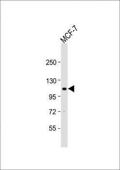
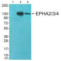
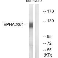
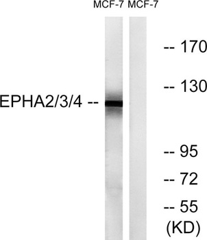
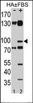
Western blot:Anti-EPHA2 Antibody (T45) at 1:1000 dilution + MCF-7.
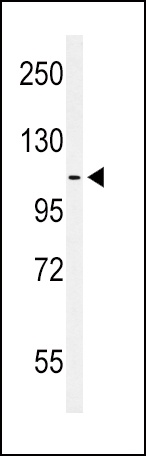
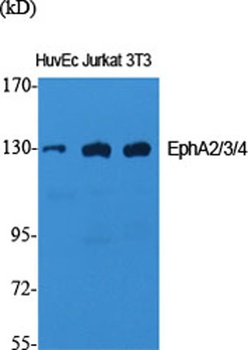
Western blot analysis of hEPHA2-T45 in MCF-7 cell line lysates. EPHA2 (arrow) was detected using the purified Pab.
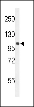
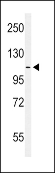
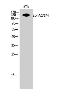
Western blot analysis of hEPHA2-T45 in mouse NIH-3T3 cell line lysates.EPHA2 (arrow) was detected using the purified Pab.
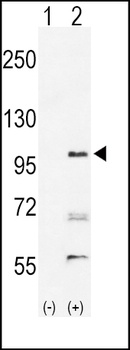
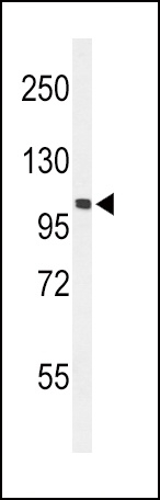
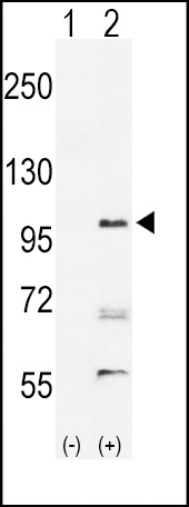
Western blot analysis of EPHA2 (arrow) using rabbit polyclonal hEPHA2-T45.293 cell lysates either nontransfected (Lane 1) or transiently transfected with the EPHA2 gene (Lane 2).
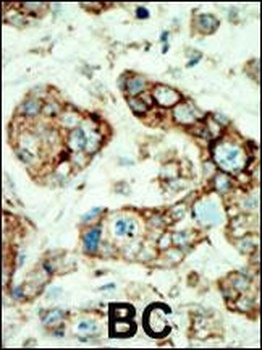
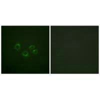
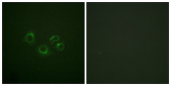
Immunohistochemical analysis of human cancer tissue reacted with the primary antibody, which was peroxidase-conjugated to the secondary antibody, followed by AEC staining.
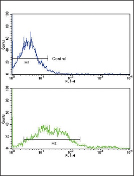
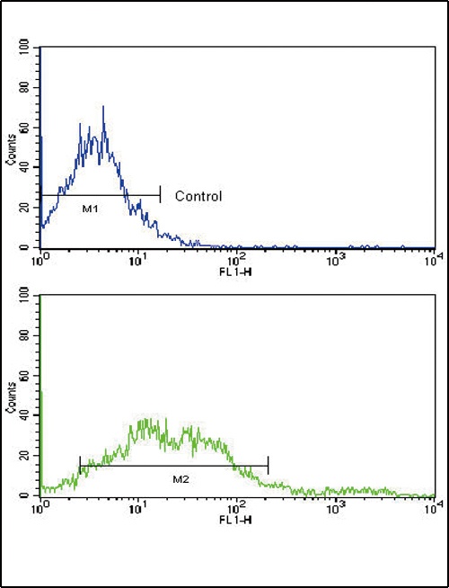
Flow cytometric analysis of NCI-H292 cells using EphA2 Antibody (N-term)(bottom histogram) compared to a negative control cell (top histogram). FITC-conjugated goat-anti-rabbit secondary antibodies were used for the analysis.
- Item 1 of 6
- Item 1 of 3
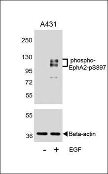
EPHA2 (Ab-588/596) antibody [orb685480]
ELISA, IF, WB
Human, Mouse
Rabbit
Polyclonal
Unconjugated
100 μl - Item 1 of 4
- Item 1 of 4
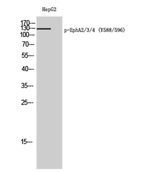
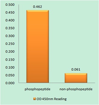
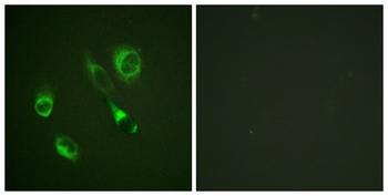
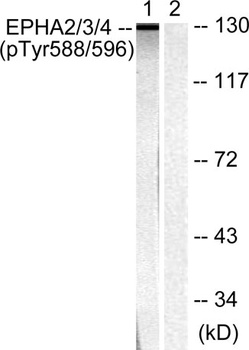
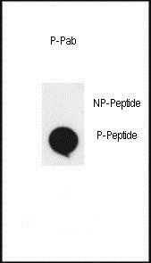
EphA2/3/4 (phospho-Tyr588/596) antibody [orb767947]
ELISA, IF, WB
Human, Mouse, Rat
Rabbit
Polyclonal
Unconjugated
50ul, 100ul - Item 1 of 3


Submit a review
Filter by Rating

- 5 stars

- 4 stars
- 3 stars
- 2 stars
- 1 stars