You have no items in your shopping cart.
Cart summary
Item 1 of 4
Item 1 of 4
EpCAM Antibody
Catalog Number: orb749655

| Catalog Number | orb749655 |
|---|---|
| Category | Antibodies |
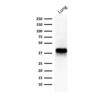
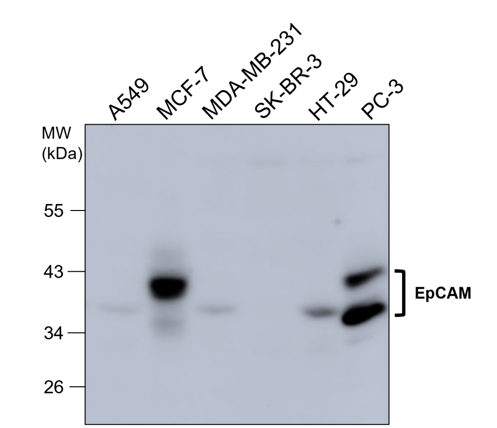
| Description | Recognizes a 40-43kDa transmembrane epithelial glycoprotein, identified as epithelial specific antigen (ESA), or epithelial cellular adhesion molecule (Ep-CAM). Ep-CAM is expressed on baso-lateral cell surface in most simple epithelia and a vast majority of carcinomas. This antibody has been used to distinguish adenocarcinoma from pleural mesothelioma and hepatocellular carcinoma. It is also useful in distinguishing serous carcinomas of the ovary from mesothelioma. This epithelial antigen plays an important role as a tumor-cell marker in lymph nodes from patients with esophageal carcinoma otherwise classified as node-negative. Epithelial antigen has also been suggested as a discriminator between basal cell and baso-squamous carcinomas, and squamous cell carcinoma of the skin. |
| Species/Host | Mouse |
| Clonality | Monoclonal |
| Clone Number | EGP40/826 |
| Tested applications | FACS, IF, IHC-P, WB |
| Reactivity | Human |
| Isotype | Mouse IgG1, kappa |
| Immunogen | A synthetic peptide (around aa 20-60) from the N-terminus of the human protein was used as the immunogen for the EpCAM antibody. |
| Dilution range | Flow cytometry: 0.5-1ug/million cells,Immunofluorescence: 1-2ug/ml,Western blot: 0.5-1ug/ml,Immunohistochemistry (FFPE): 0.5-1ug/ml for 30 min at RT |
| Purity | Protein G affinity chromatography |
| Conjugation | Unconjugated |
| Formula | 0.2 mg/ml in 1X PBS with 0.1 mg/ml BSA (US sourced) and 0.05% sodium azide |
| Hazard Information | This EpCAM antibody is available for research use only. |
| UniProt ID | P16422 |
| Storage | Store the EpCAM antibody at 2-8°C (with azide) or aliquot and store at -20°C or colder (without azide). |
| Buffer/Preservatives | 0.2 mg/ml in 1X PBS with 0.1 mg/ml rAlbumin (US sourced) and 0.05% sodium azide |
| Note | For research use only |
| Application notes | Optimal dilution of the EpCAM antibody should be determined by the researcher.1. Staining of formalin-fixed tissues requires boiling tissue sections in 10mM Citrate buffer, pH 6.0, for 10-20 min followed by cooling at RT for 20 min2. The prediluted format is supplied in a dropper bottle and is optimized for use in IHC. After epitope retrieval step (if required), drip mAb solution onto the tissue section and incubate at RT for 30 min. |
| Expiration Date | 12 months from date of receipt. |

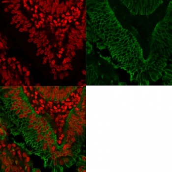
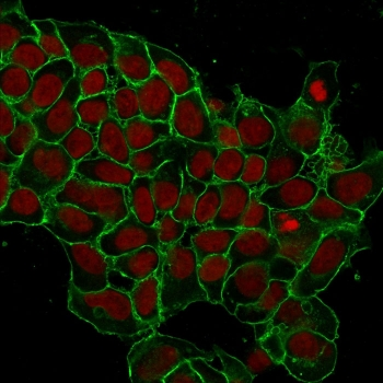
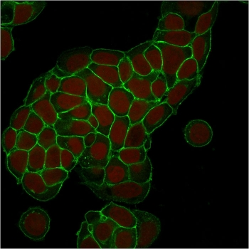
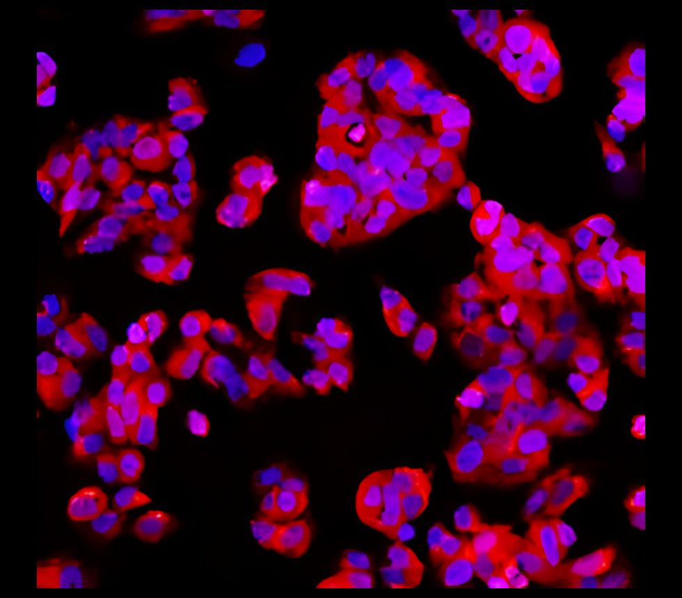
Right: Confocal Immunofluorescent analysis of SK-OV-3 cells using AF488-labeled EpCAM antibody (EGP40/826) (Green). F-actin filaments were labeled with DyLight 554 Phalloidin (red). Left: Negative control. DAPI was used to stain the cell nuclei (blue).

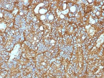
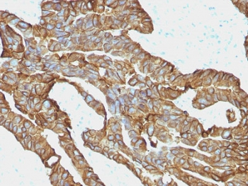
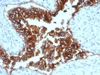
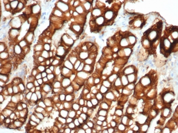
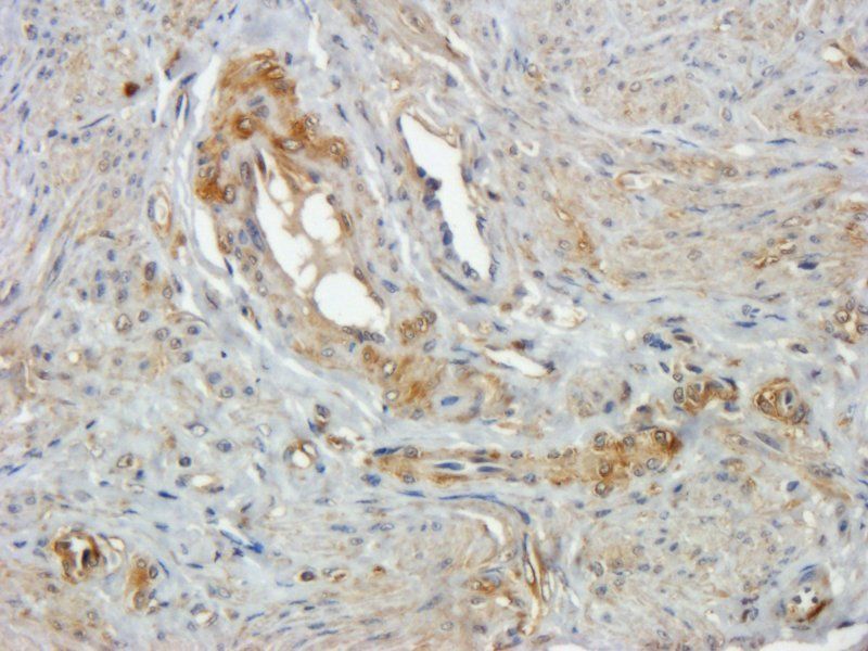
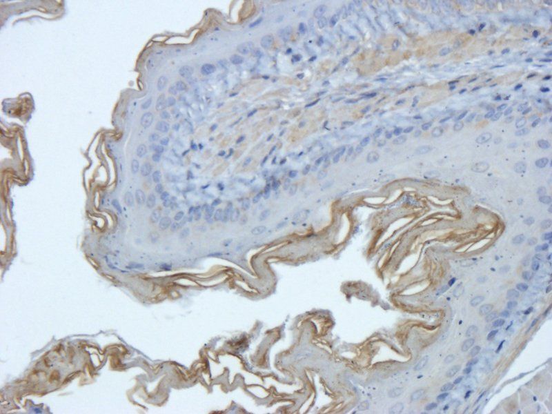
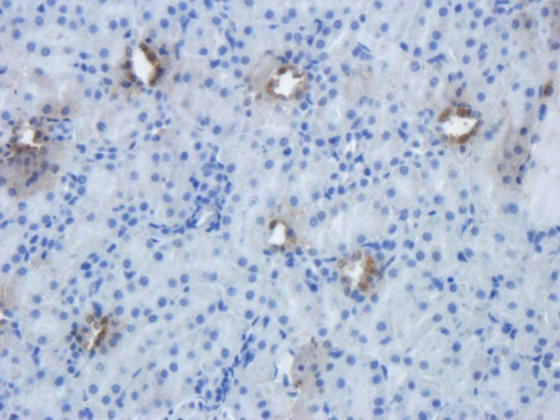
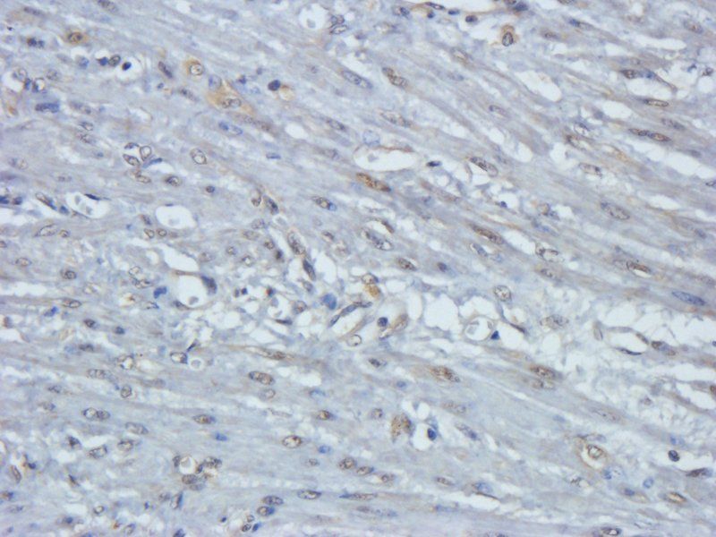
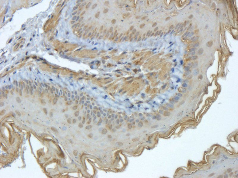
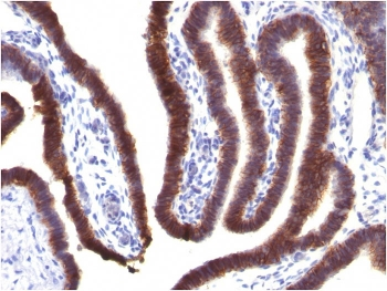
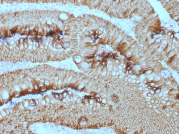
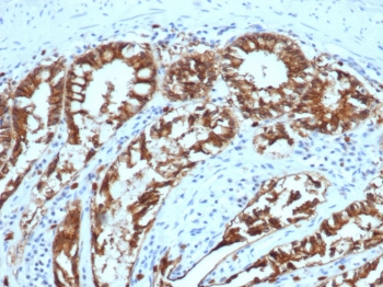
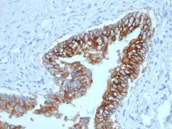
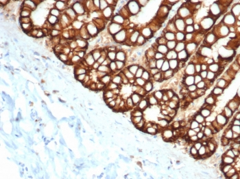
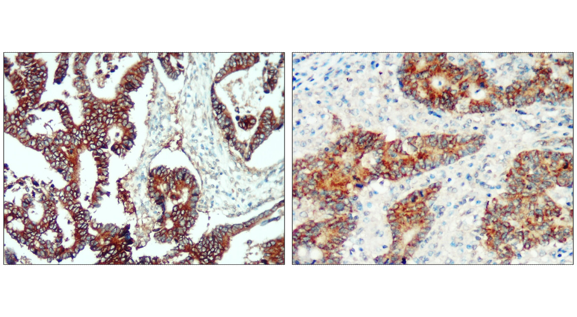
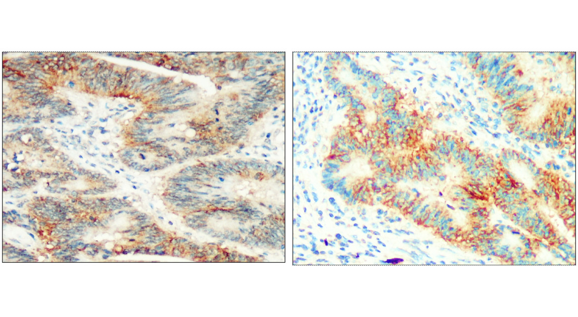
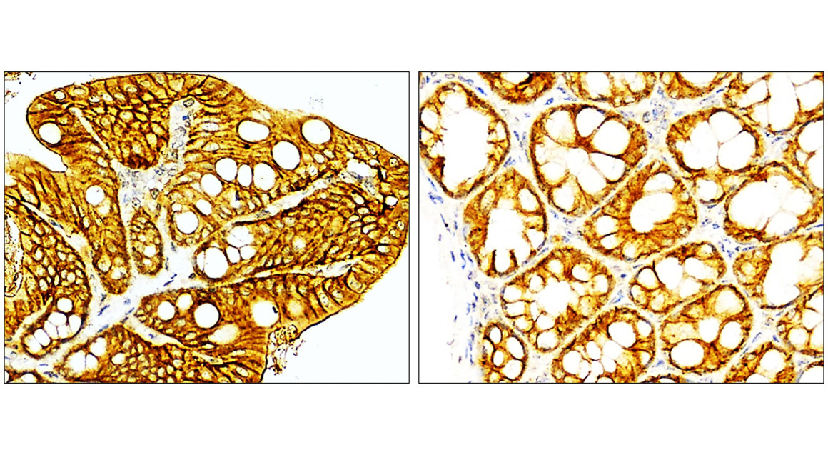
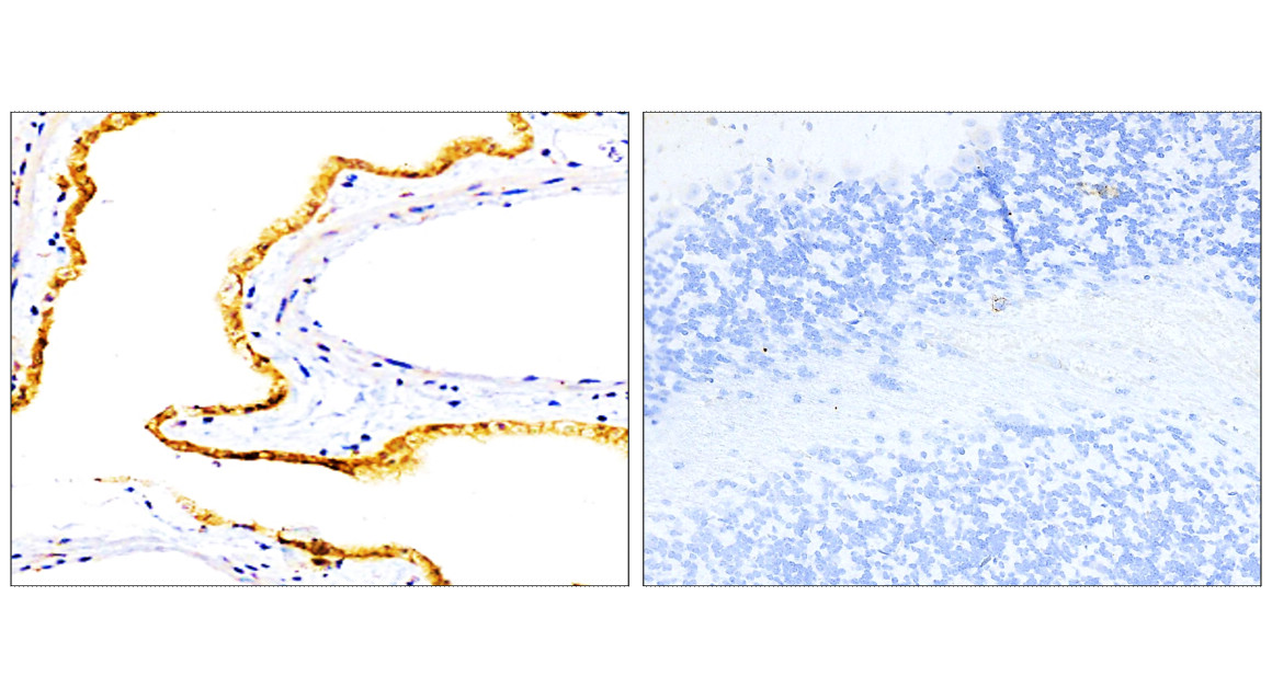
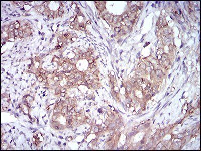
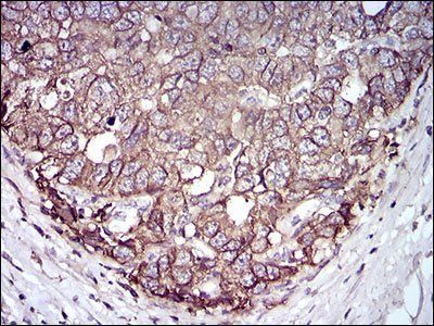
IHC: Formalin-fixed, paraffin-embedded human breast carcinoma stained with EpCAM antibody (EGP40/826).

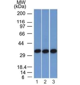
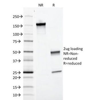
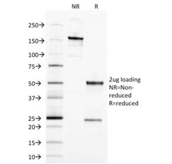
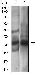
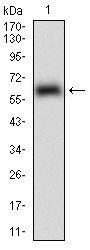
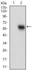
SDS-PAGE Analysis of Purified, BSA-Free EpCAM Antibody (clone EGP40/826). Confirmation of Integrity and Purity of the Antibody.

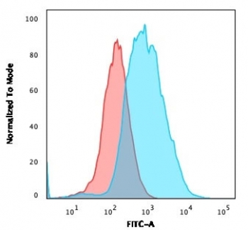
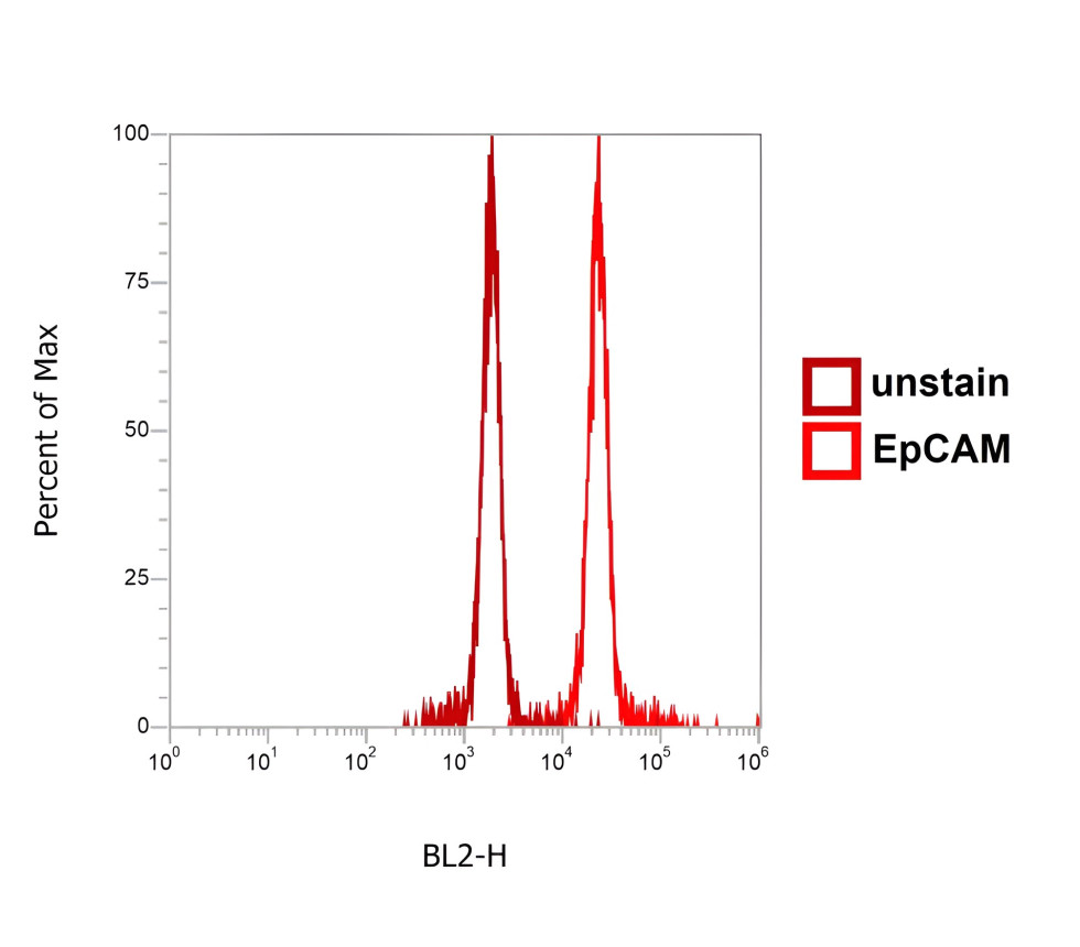
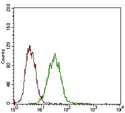
Flow cytometry testing of PFA-fixed human MCF7 cells with EpCAM antibody (clone EGP40/826); Red=isotype control, Blue=EpCAM antibody.
- Item 1 of 11
EpCAM Antibody / Extracellular domain [orb606357]
FACS, IF, IHC-P, WB
Canine, Feline, Human
Mouse
Monoclonal
Unconjugated
20 μg, 100 μg - Item 1 of 5
- Item 1 of 9
EpCAM Antibody [orb639787]
IF, IHC-P, WB
Canine, Feline, Human
Mouse
Monoclonal
Unconjugated
20 μg, 100 μg - Item 1 of 8
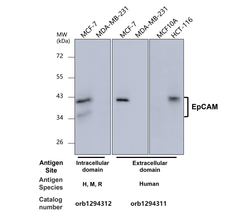
EpCAM (Extracellular domain) antibody [orb1294311]
IF, IHC, WB
Human
Rabbit
Polyclonal
Unconjugated
100 μl, 25 μl - Item 1 of 7

Submit a review
Filter by Rating

- 5 stars

- 4 stars
- 3 stars
- 2 stars
- 1 stars
