You have no items in your shopping cart.
Cart summary
Item 1 of 4
Item 1 of 4
EGR1 Antibody (N-term)
Catalog Number: orb1931351

| Catalog Number | orb1931351 |
|---|---|
| Category | Antibodies |
| Description | Purified Rabbit Polyclonal Antibody (Pab) |
| Target | This EGR1 antibody is generated from rabbits immunized with a KLH conjugated synthetic peptide between 9-37 amino acids from the N-terminal region of human EGR1. |
| Clonality | Polyclonal |
| Species/Host | Rabbit |
| Isotype | Rabbit IgG |
| Conjugation | Unconjugated |
| Reactivity | Human, Mouse |
| Predicted Reactivity | Other |
| Form/Appearance | Purified polyclonal antibody supplied in PBS with 0.09% (W/V) sodium azide. This antibody is prepared by Saturated Ammonium Sulfate (SAS) precipitation followed by dialysis against PBS. |
| UniProt ID | P18146 |
| MW | 57507 Da |
| Tested applications | FC, IF, WB |
| Dilution range | IF: 1:10~50, WB: 1:1000, WB: 1:1000, FC: 1:10~50 |
| Antibody Type | Primary Antibody |
| Clone Number | RB19179 |
| Storage | Maintain refrigerated at 2-8°C for up to 2 weeks. For long term storage store at -20°C in small aliquots to prevent freeze-thaw cycles |
| Alternative names | Early growth response protein 1, EGR-1, AT225, Ner Read more... |
| Note | For research use only |
| NCBI | NP_001955.1 |
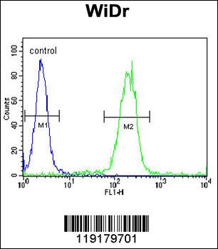
EGR1 Antibody (N-term) flow cytometric analysis of WiDr cells (right histogram) compared to a negative control cell (left histogram). FITC-conjugated goat-anti-rabbit secondary antibodies were used for the analysis.
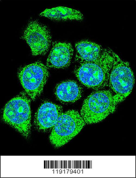
Confocal immunofluorescent analysis of EGR1 Antibody (N-term) with hela cell followed by Alexa Fluor 488-conjugated goat anti-rabbit lgG (green). DAPI was used to stain the cell nuclear (blue).
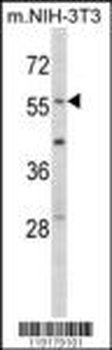
Western blot analysis of EGR1 Antibody (N-term) in mouse NIH-3T3 tissue lysates (35 ug/lane). EGR1 (arrow) was detected using the purified Pab.
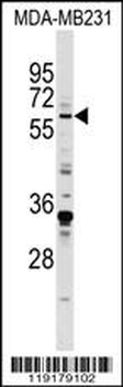
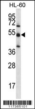
EGR1 Antibody (N-term) western blot analysis in MDA-MB231 cell line lysates (35 ug/lane). This demonstrates the EGR1 antibody detected the EGR1 protein (arrow).
- Item 1 of 1