You have no items in your shopping cart.
Cart summary
Item 1 of 3
Item 1 of 3
EGFR Antibody
Catalog Number: orb623853
| Catalog Number | orb623853 |
|---|---|
| Category | Antibodies |
| Description | EGFR Antibody |
| Species/Host | Rabbit |
| Clonality | Polyclonal |
| Tested applications | ELISA, FC, IHC, WB |
| Reactivity | Mouse, Rat |
| Isotype | Rabbit IgG |
| Immunogen | E.coli-derived mouse EGFR recombinant protein (Position: L25-P596). |
| Concentration | Adding 0.2 ml of distilled water will yield a concentration of 500 μg/ml. |
| Dilution range | Western blot, 0.25-0.5μg/ml, Human, Mouse, Rat Immunohistochemistry (Paraffin-embedded Section), 0.5-1μg/ml, Human, Mouse, Rat Flow Cytometry, 1-3μg/1x106 cells, Human Direct ELISA, 0.1-0.5μg/ml, Human |
| Form/Appearance | Lyophilized |
| Conjugation | Unconjugated |
| MW | 180 kDa |
| UniProt ID | Q01279 |
| Storage | Store at -20˚C for one year from date of receipt. After reconstitution, at 4˚C for one month. It can also be aliquotted and stored frozen at -20˚C for six months. Avoid repeated freeze-thaw cycles. |
| Alternative names | Epidermal growth factor receptor; Egfr Read more... |
| Note | For research use only |
| Application notes | Tested Species: In-house tested species with positive results. Other applications have not been tested. Optimal dilutions should be determined by end users. Add 0.2ml of distilled water will yield a concentration of 500ug/ml. |
| Expiration Date | 12 months from date of receipt. |
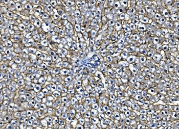
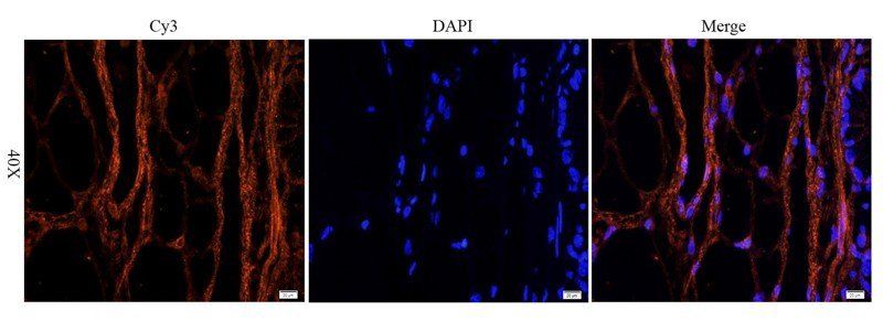
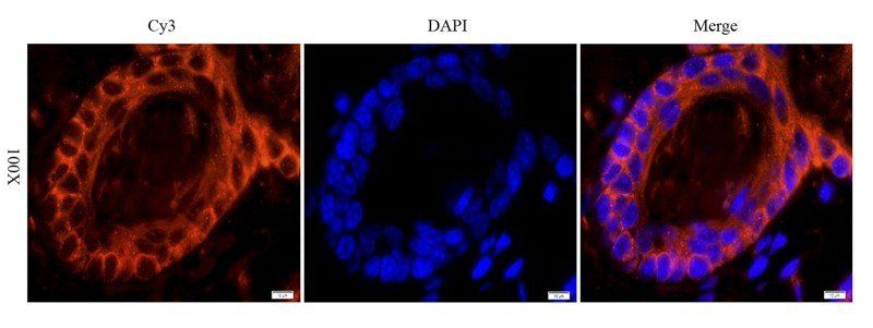
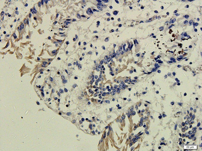
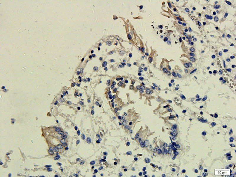
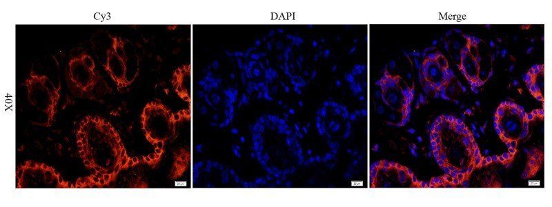
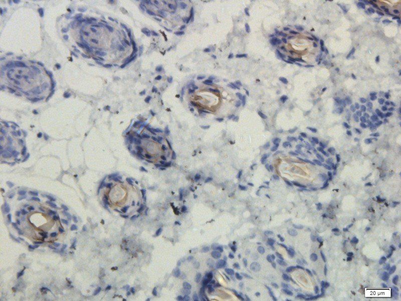
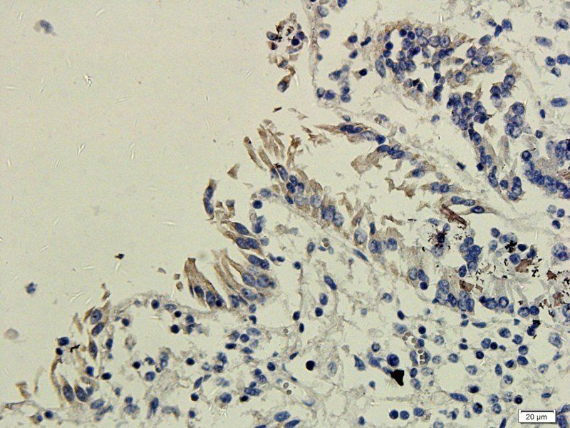
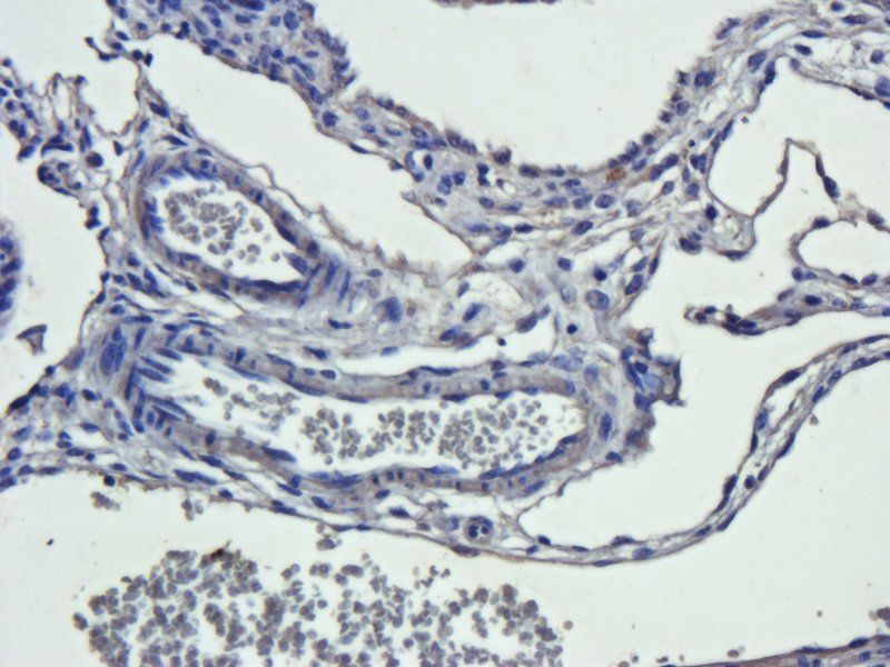
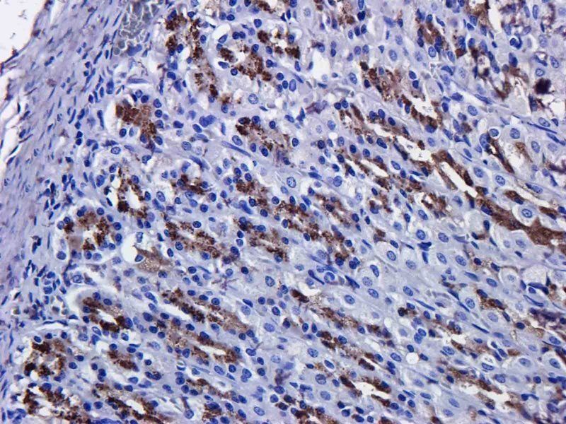
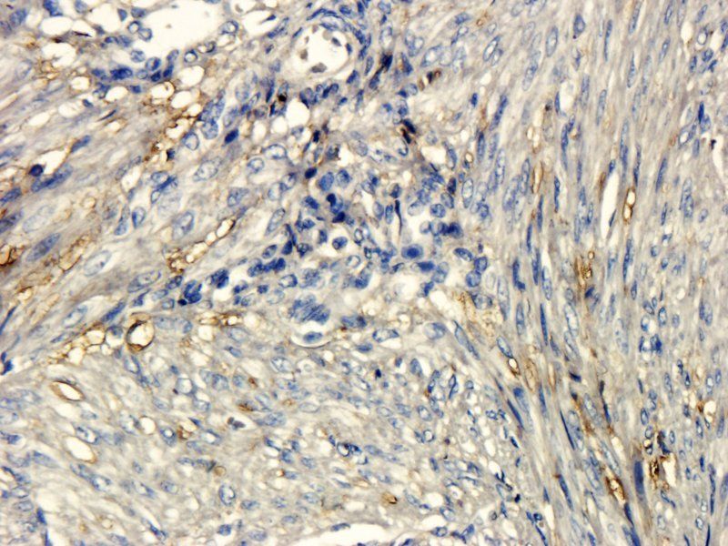
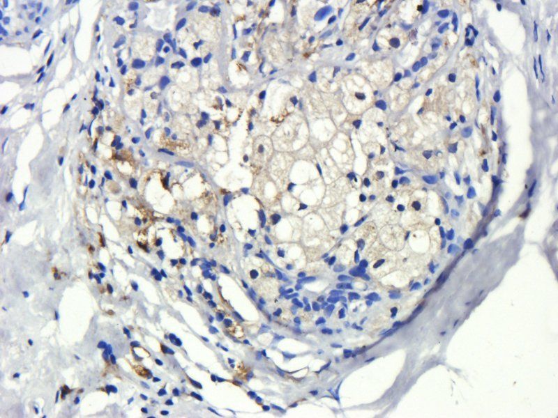
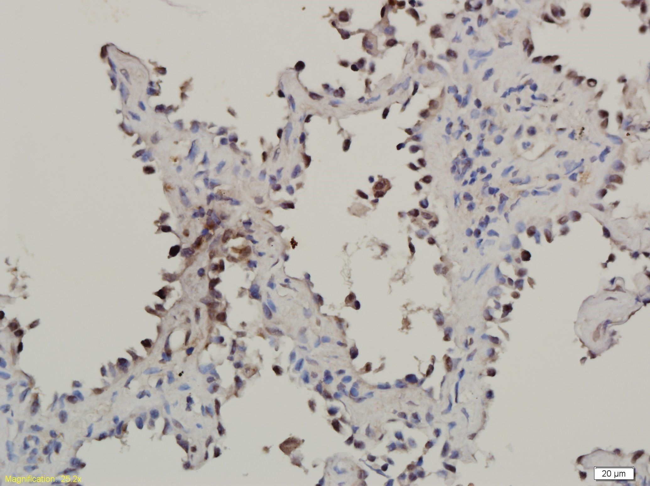
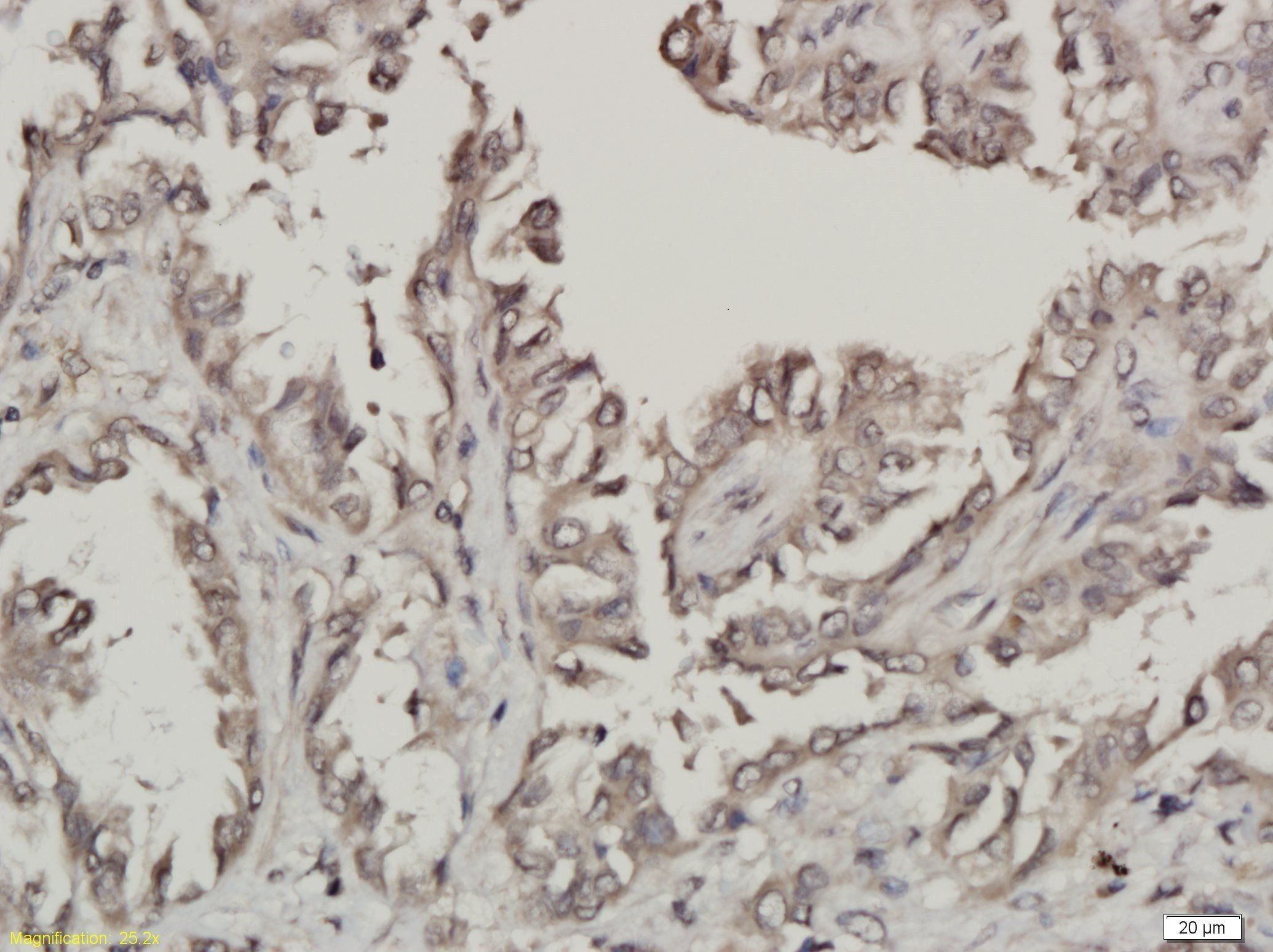
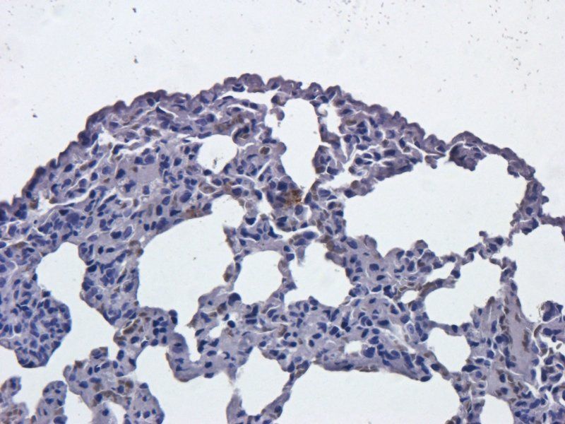
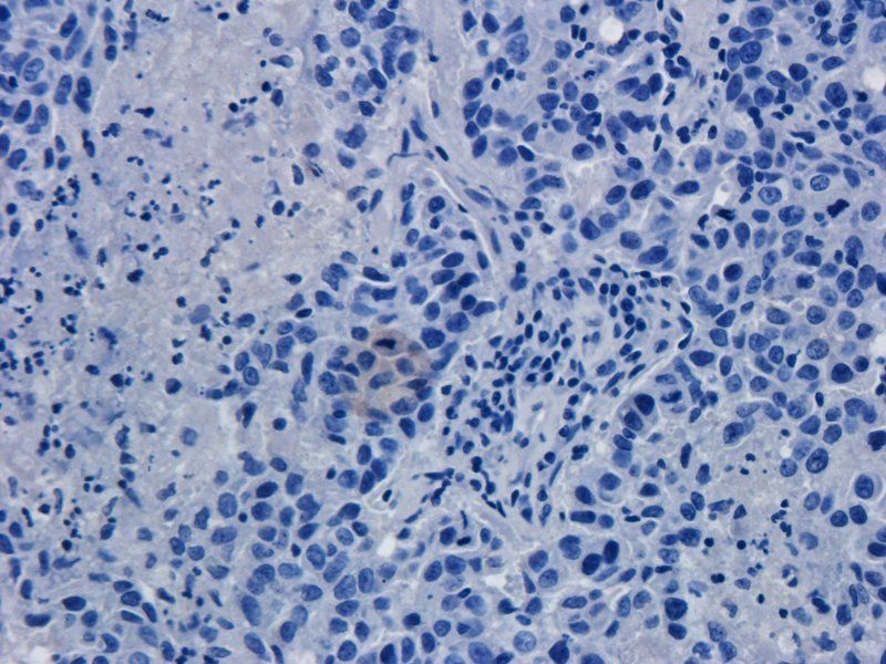
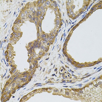
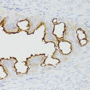
IHC analysis of EGFR using anti-EGFR antibody (orb623853). EGFR was detected in paraffin-embedded section of mouse liver tissue. Heat mediated antigen retrieval was performed in EDTA buffer (pH8.0, epitope retrieval solution). The tissue section was blocked with 10% goat serum. The tissue section was then incubated with 1μg/ml rabbit anti-EGFR Antibody (orb623853) overnight at 4°C. Biotinylated goat anti-rabbit IgG was used as secondary antibody and incubated for 30 minutes at 37°C. The tissue section was developed using Strepavidin-Biotin-Complex (SABC) (Catalog # orb90444) with DAB as the chromogen.
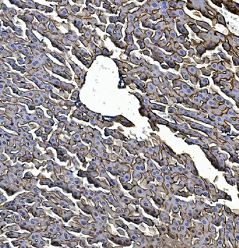
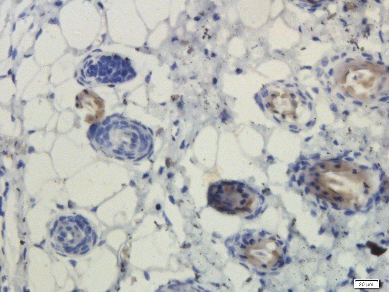
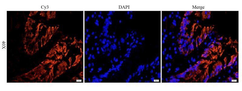
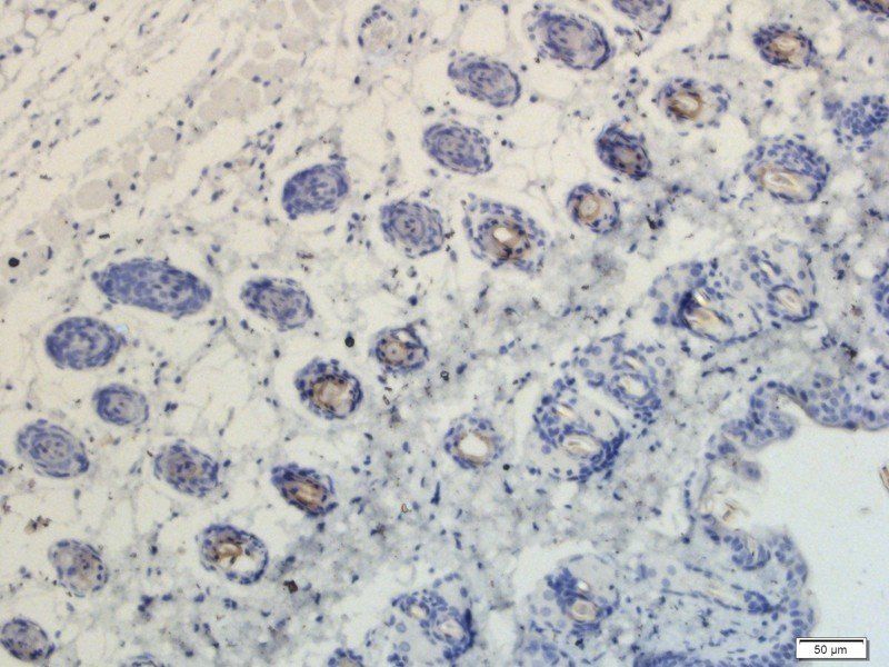
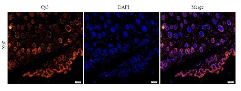
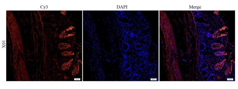
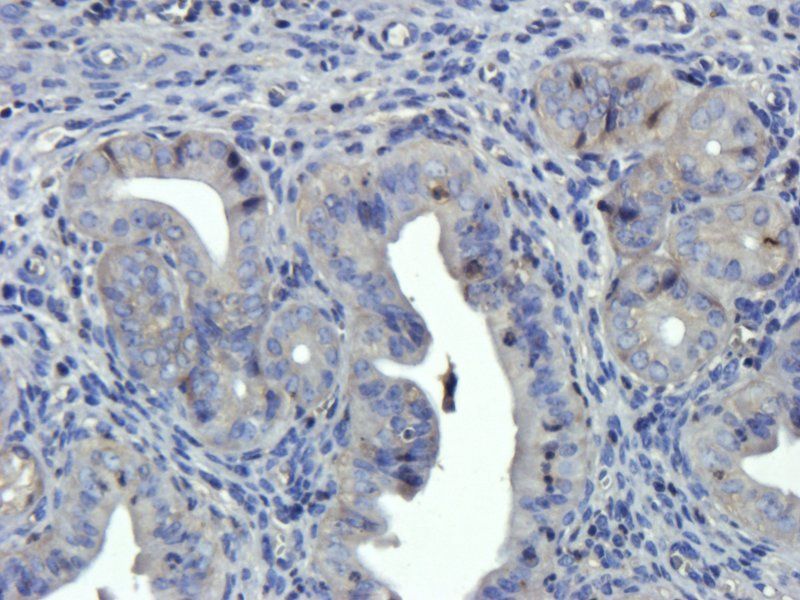
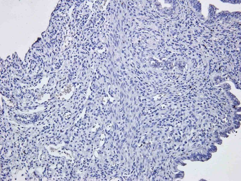
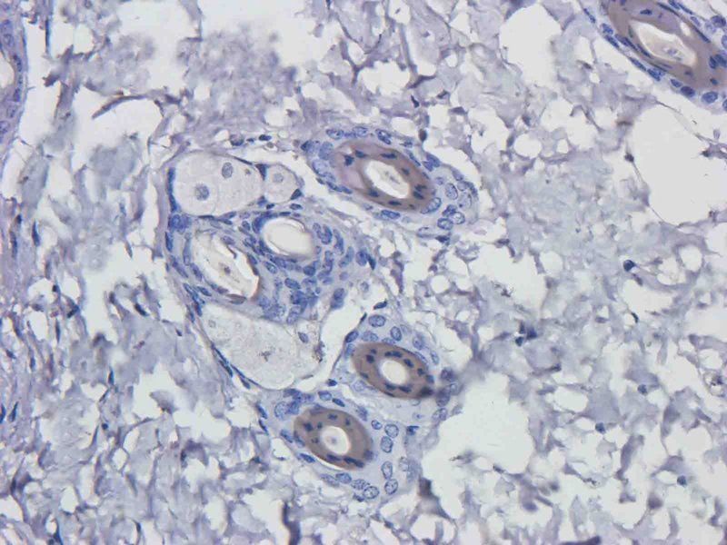
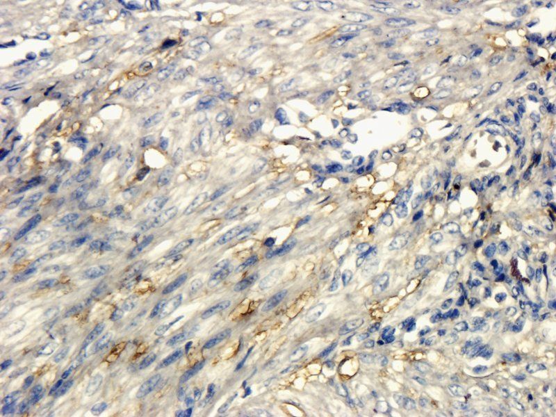
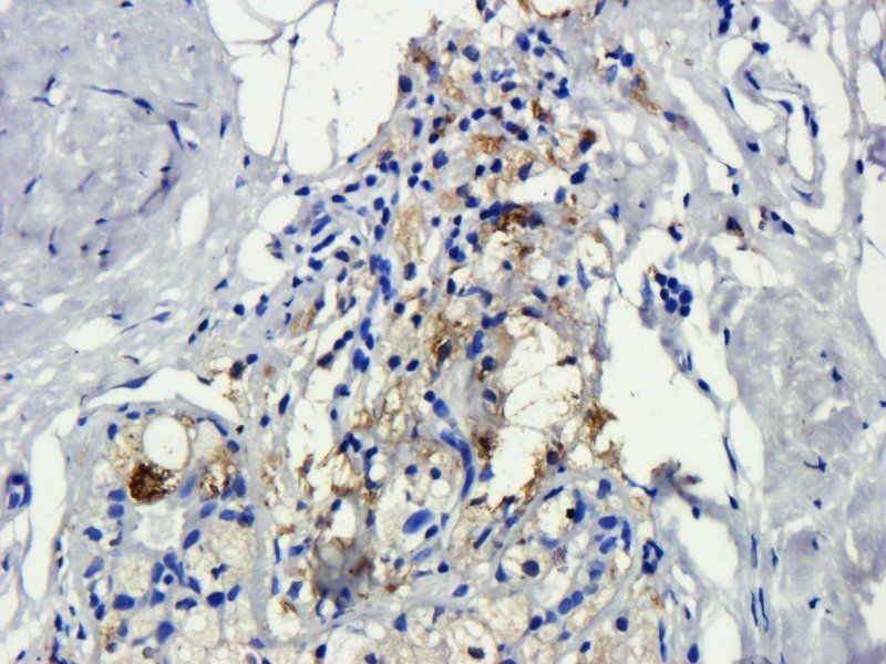
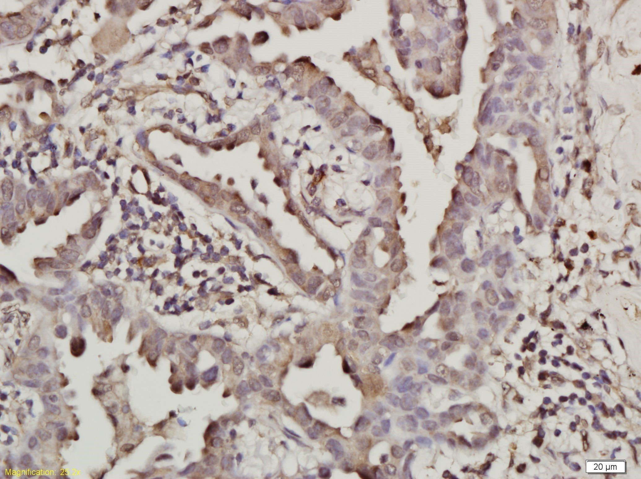
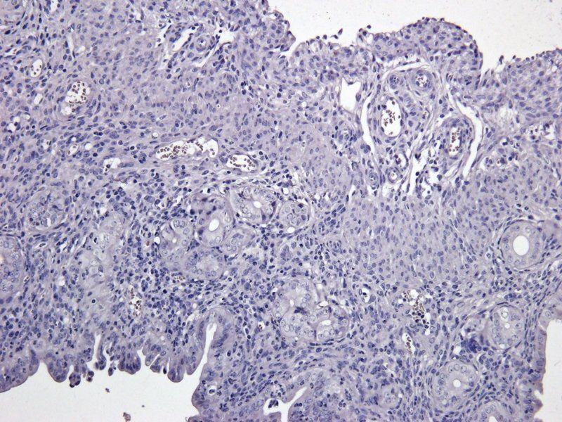
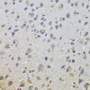
IHC analysis of EGFR using anti-EGFR antibody (orb623853). EGFR was detected in paraffin-embedded section of rat liver tissue. Heat mediated antigen retrieval was performed in EDTA buffer (pH8.0, epitope retrieval solution). The tissue section was blocked with 10% goat serum. The tissue section was then incubated with 1μg/ml rabbit anti-EGFR Antibody (orb623853) overnight at 4°C. Biotinylated goat anti-rabbit IgG was used as secondary antibody and incubated for 30 minutes at 37°C. The tissue section was developed using Strepavidin-Biotin-Complex (SABC) (Catalog # orb90444) with DAB as the chromogen.
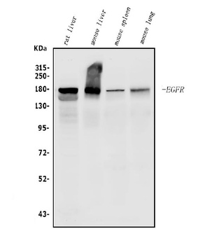
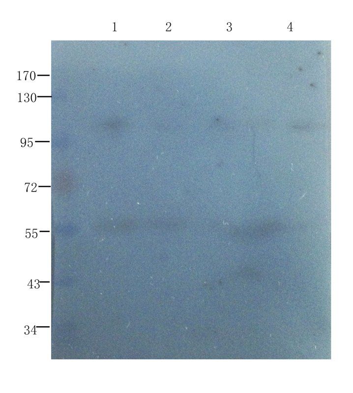
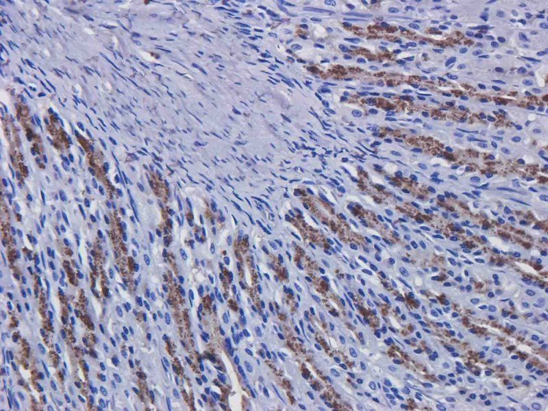
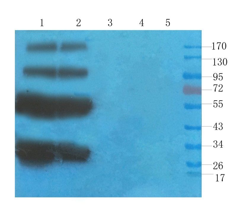
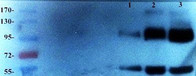
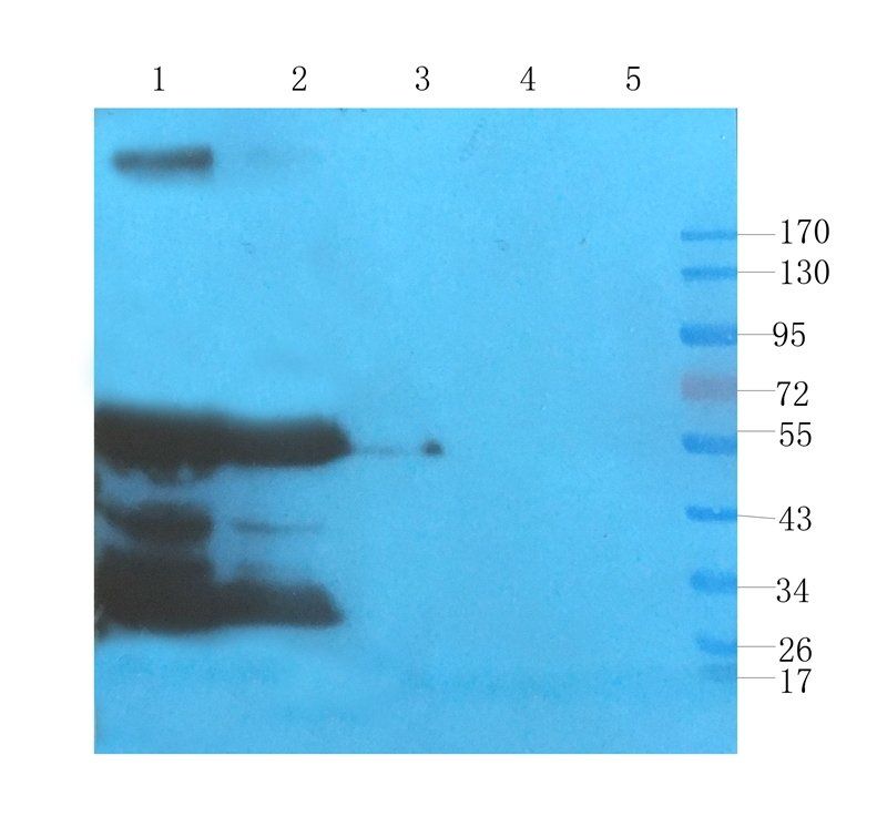
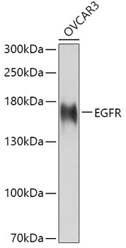
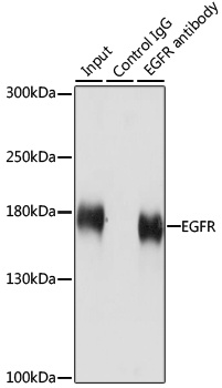
Western blot analysis of EGFR using anti-EGFR antibody (orb623853). Electrophoresis was performed on a 5-20% SDS-PAGE gel at 70V (Stacking gel)/90V (Resolving gel) for 2-3 hours. The sample well of each lane was loaded with 50ug of sample under reducing conditions. Lane 1: rat liver tissue lysates, Lane 2: mouse liver tissue lysates, Lane 3: mouse spleen tissue lysates, Lane 4: mouse lung tissue lysates. After Electrophoresis, proteins were transferred to a Nitrocellulose membrane at 150mA for 50-90 minutes. Blocked the membrane with 5% Non-fat Milk/TBS for 1.5 hour at RT. The membrane was incubated with rabbit anti-EGFR antigen affinity purified polyclonal antibody (Catalog # orb623853) at 0.5 μg/mL overnight at 4°C, then washed with TBS-0.1%Tween 3 times with 5 minutes each and probed with a goat anti-rabbit IgG-HRP secondary antibody at a dilution of 1:5000 for 1.5 hour at RT. The signal is developed using an Enhanced Chemiluminescent detection (ECL) kit (Catalog # orb90503) with Tanon 5200 system. A specific band was detected for EGFR at approximately 180KD. The expected band size for EGFR is at 180KD.
- Item 1 of 18
EGFR isoform a variant antibody [orb308736]
ELISA, ICC, IF, IHC-P, WB
Human, Mouse, Porcine, Rat
Rabbit
Polyclonal
Unconjugated
100 μg, 200 μg - Item 1 of 8
- Item 1 of 9
- Item 1 of 7
- Item 1 of 8

Submit a review
Filter by Rating

- 5 stars

- 4 stars
- 3 stars
- 2 stars
- 1 stars