You have no items in your shopping cart.
Cart summary
Item 1 of 3
Item 1 of 3
EGFR antibody
Catalog Number: orb304743

| Catalog Number | orb304743 |
|---|---|
| Category | Antibodies |
| Description | Rabbit polyclonal antibody to EGFR which is overexpressed in a variety of human epithelial tumors, often as a consequence of gene amplification. Tumors with EGFR gene amplification frequently contain EGFR gene rearrangements, with the most common extracellular domain mutation being EGFRvIII. |
| Species/Host | Rabbit |
| Clonality | Polyclonal |
| Tested applications | IF, IH, WB |
| Reactivity | Human, Mouse, Porcine, Rat, Zebrafish |
| Immunogen | KLH-conjugated synthetic peptide encompassing a sequence within the center region of human EGFR. The exact sequence is proprietary. |
| Dilution range | WB: 1:500-1000, IHC-P: 1:100-200, IF/ICC: 1:100-500 |
| Form/Appearance | Liquid in 0.42% Potassium phosphate, 0.87% Sodium chloride, pH 7.3, 30% glycerol, and 0.01% sodium azide. |
| Conjugation | Unconjugated |
| Target | EGFR |
| Entrez | 13649, 1956 |
| UniProt ID | P00533, Q01279 |
| Source | Rabbit |
| Storage | Shipped at 4°C. Upon delivery aliquot and store at -20°C for one year. Avoid freeze/thaw cycles. |
| Buffer/Preservatives | Liquid in 0.42% Potassium phosphate, 0.87% Sodium chloride, pH 7.3, 30% glycerol, and 0.01% sodium azide. |
| Alternative names | anti-ERBB antibody, anti-ERBB1 antibody, anti-HER1 Read more... |
| Note | For research use only |
| Expiration Date | 12 months from date of receipt. |
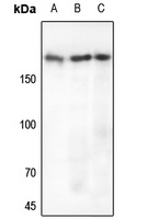
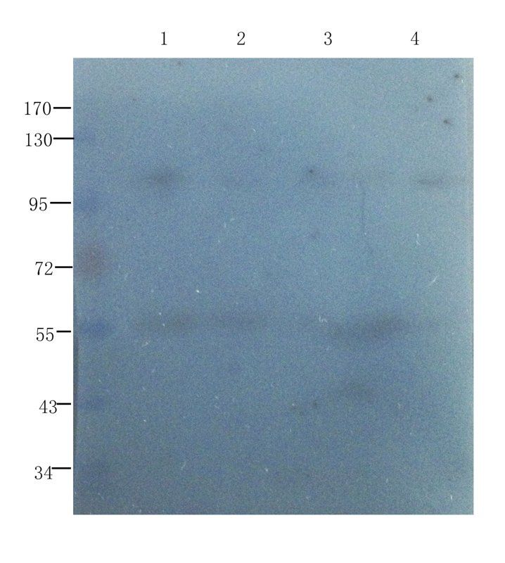
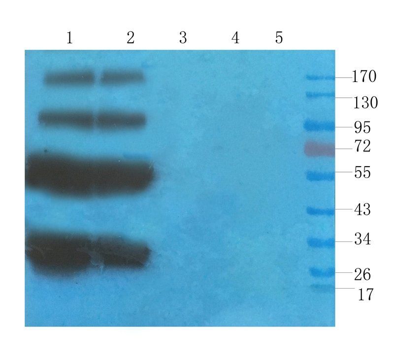
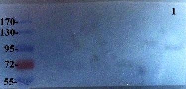
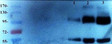
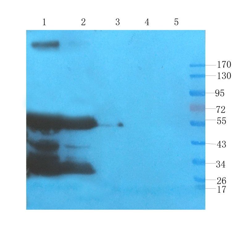
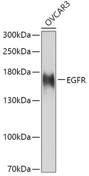
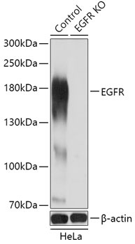
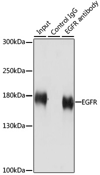
Western blot analysis of EGFR expression in A549 (A), mouse liver (B), rat liver (C) whole cell lysates. (Predicted band size: 134 kD; Observed band size: 175 kD)
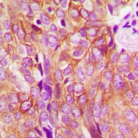
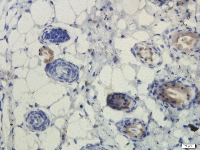
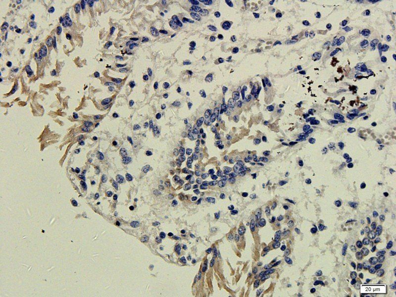
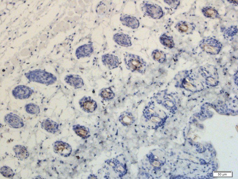
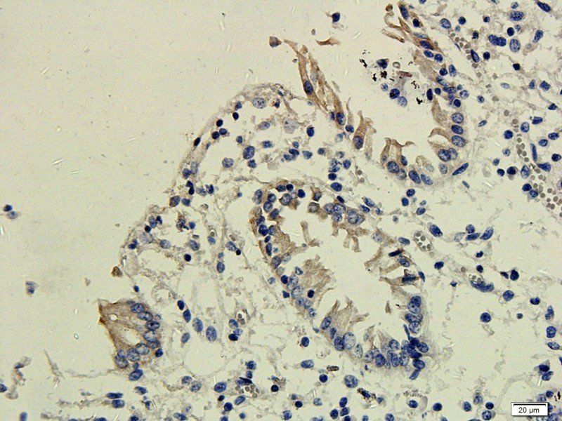
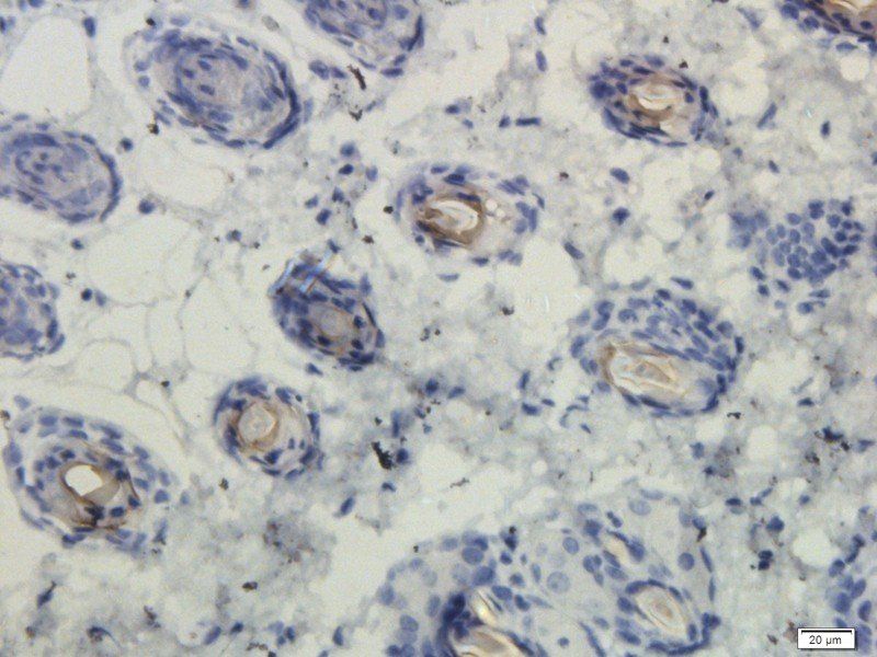
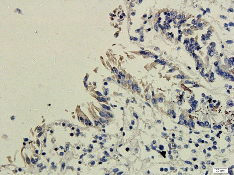
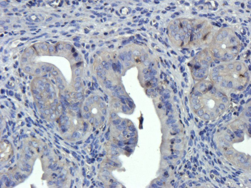
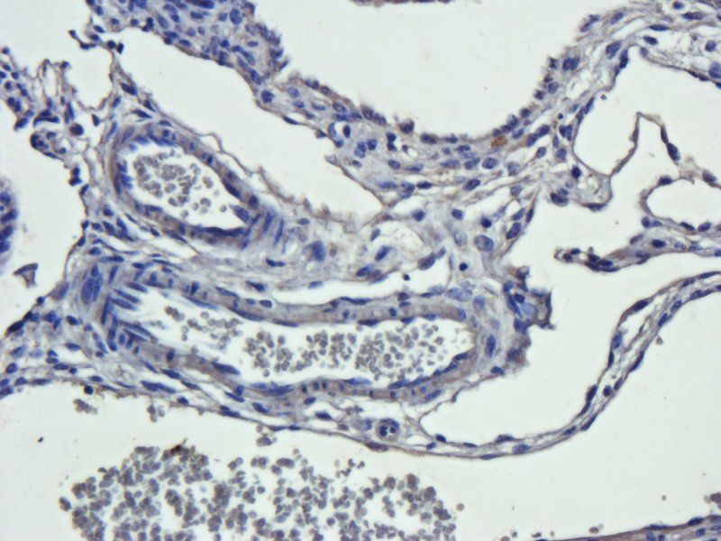
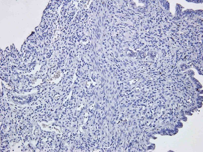
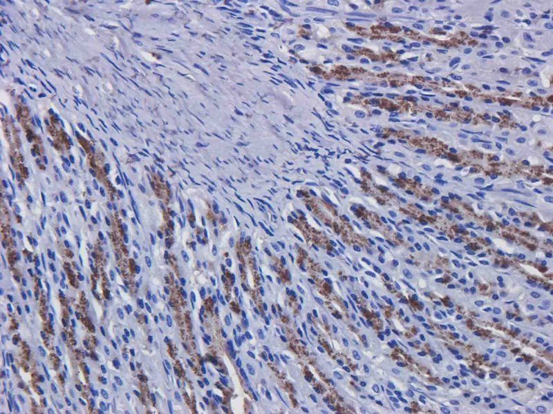
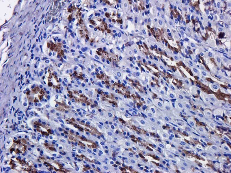
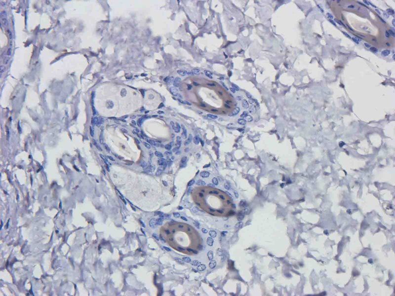
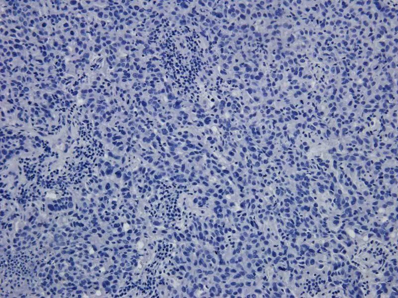
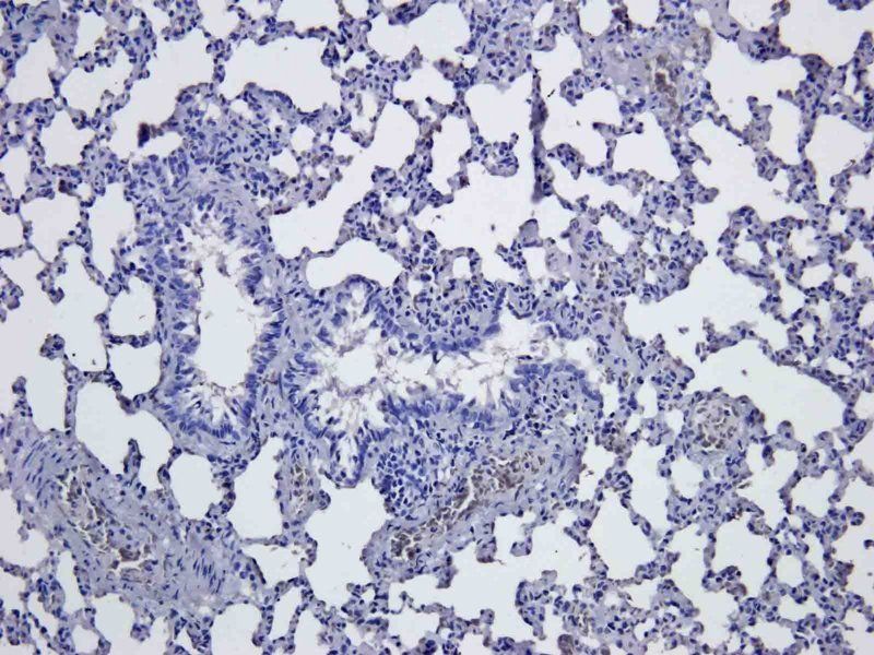
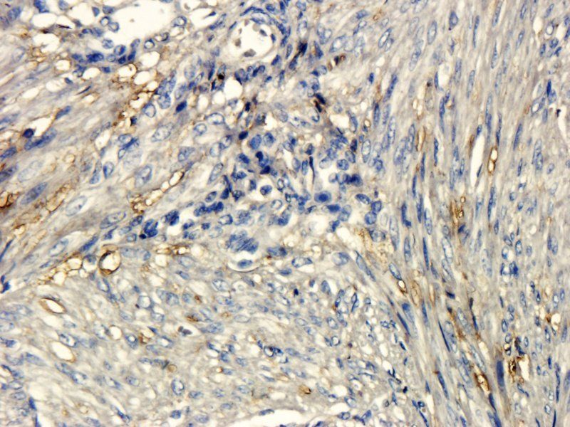
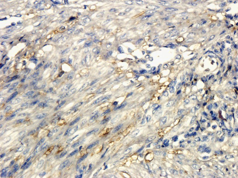
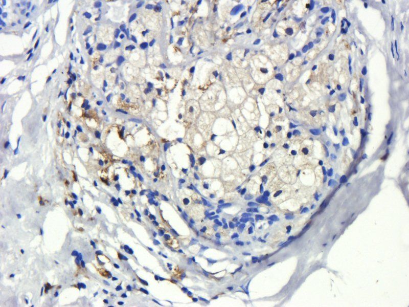
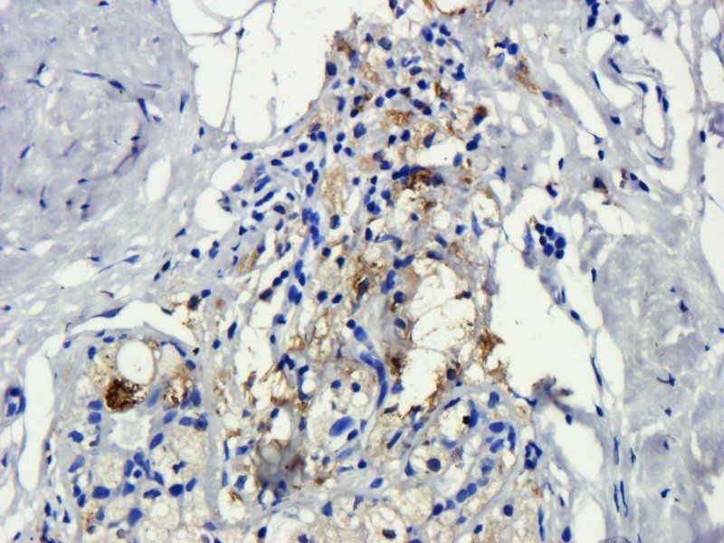
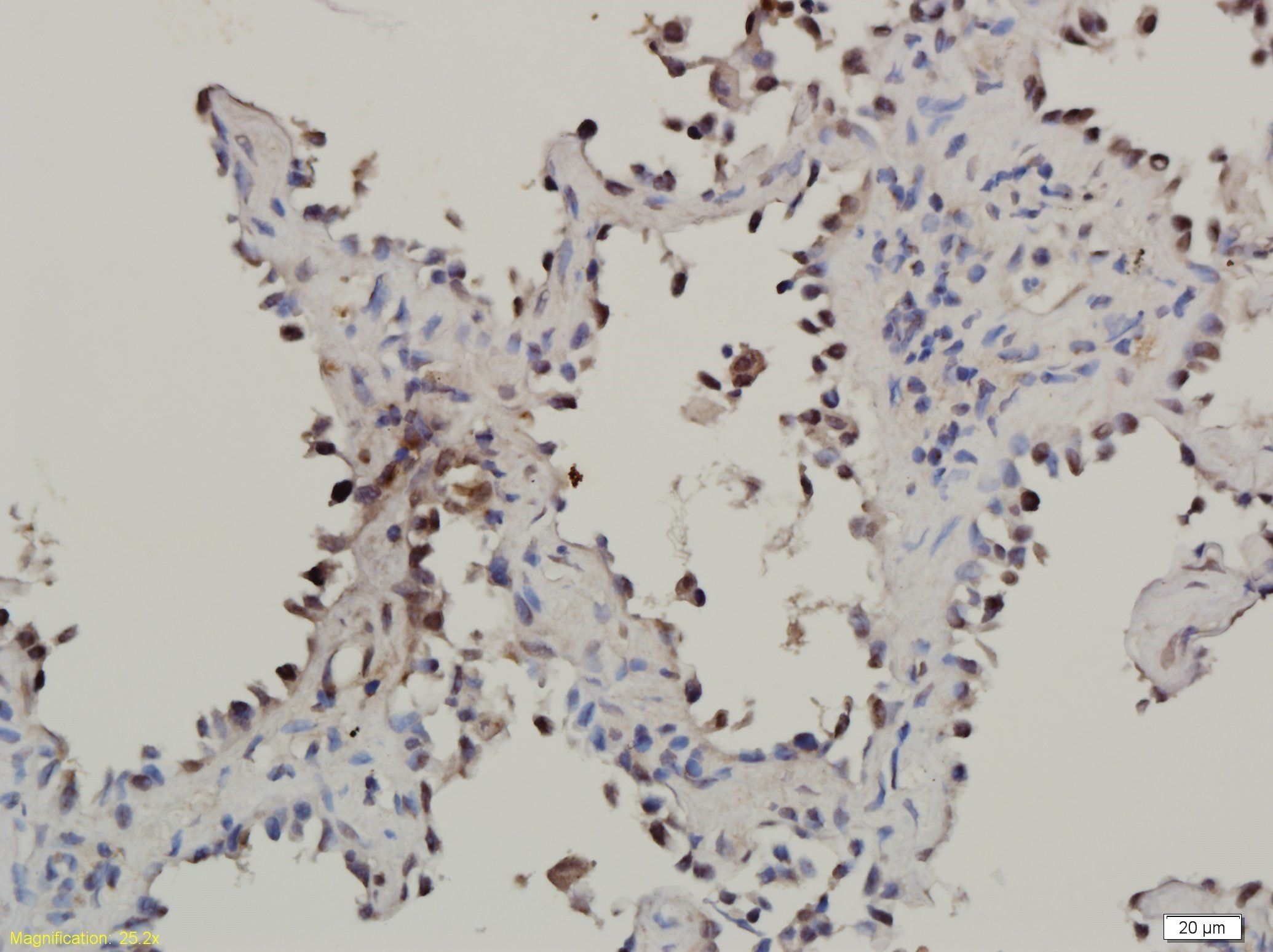
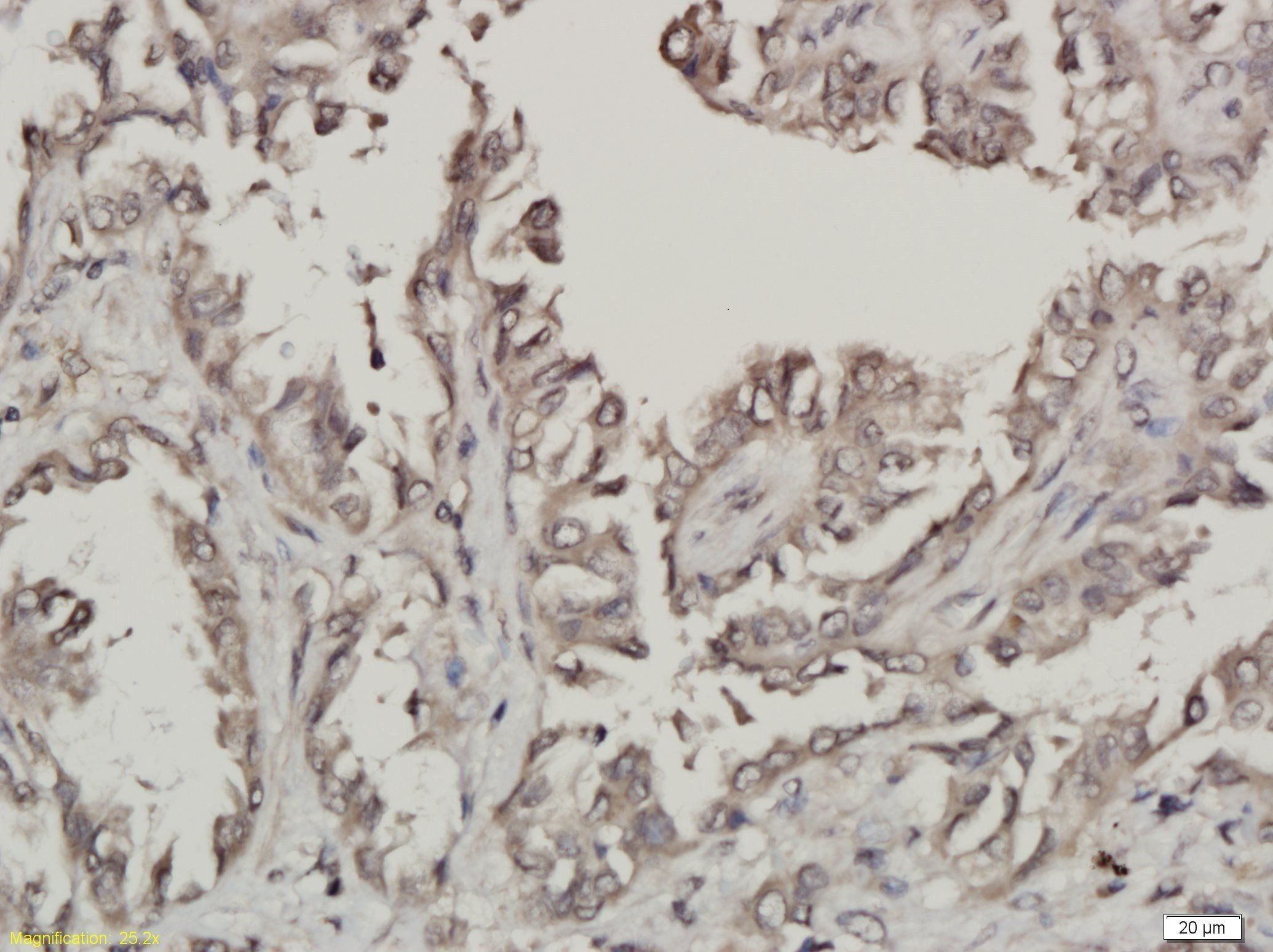
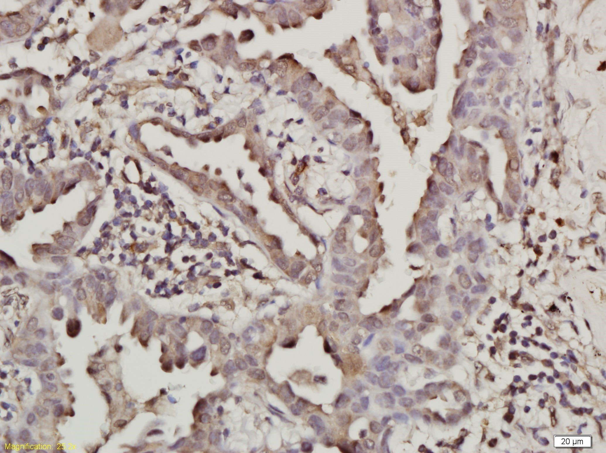
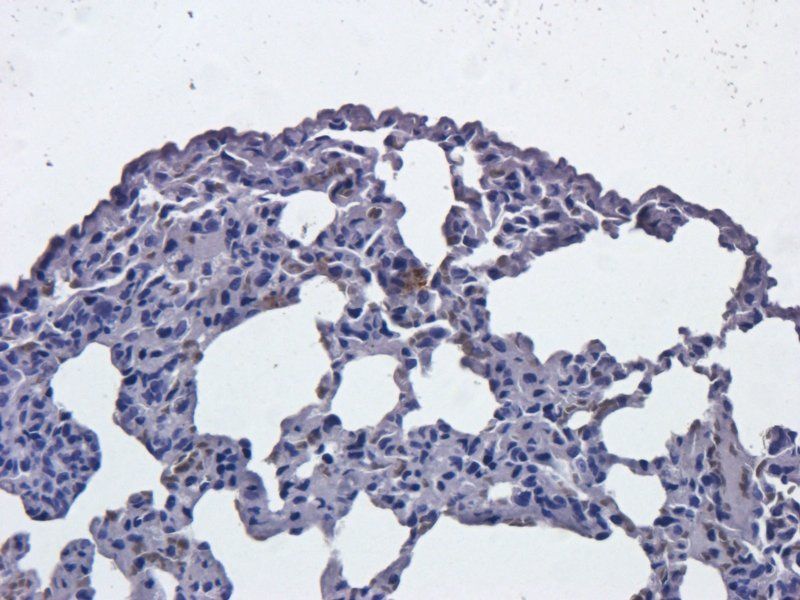
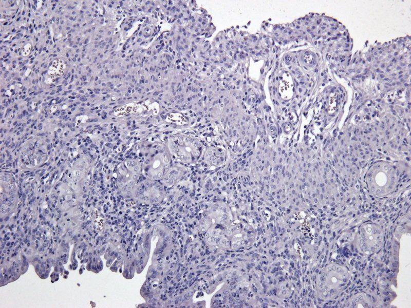
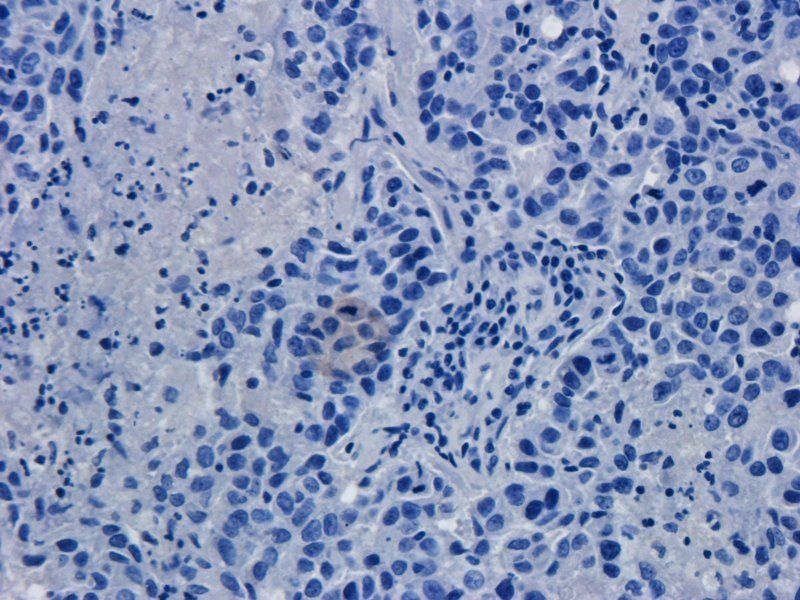
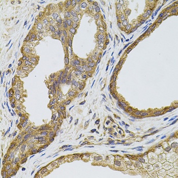
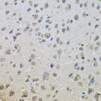
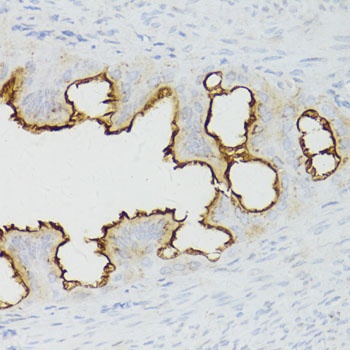
Immunohistochemical analysis of EGFR staining in human breast cancer formalin fixed paraffin embedded tissue section. The section was pre-treated using heat mediated antigen retrieval with sodium citrate buffer (pH 6.0). The section was then incubated with the antibody at room temperature and detected using an HRP conjugated compact polymer system. DAB was used as the chromogen. The section was then counterstained with haematoxylin and mounted with DPX.
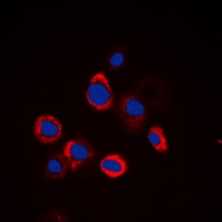
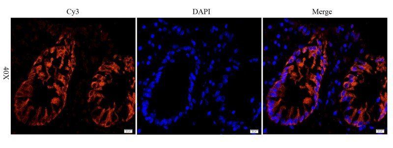
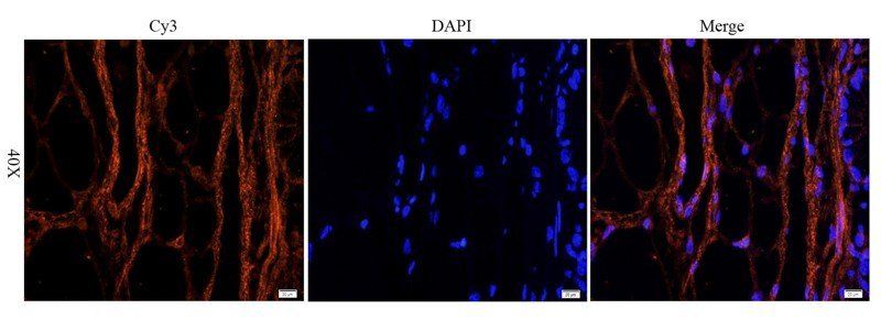
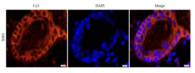
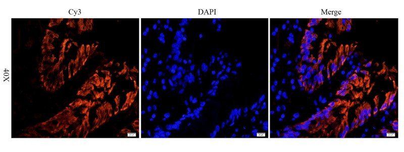
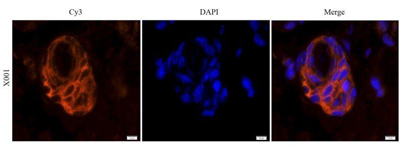
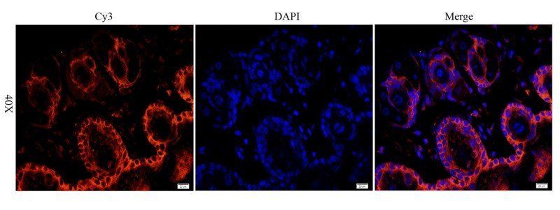
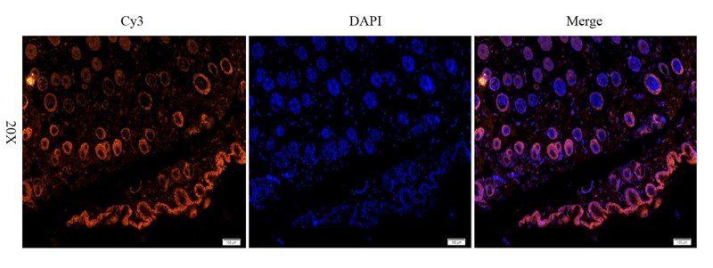
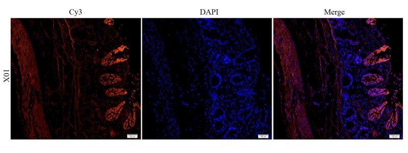
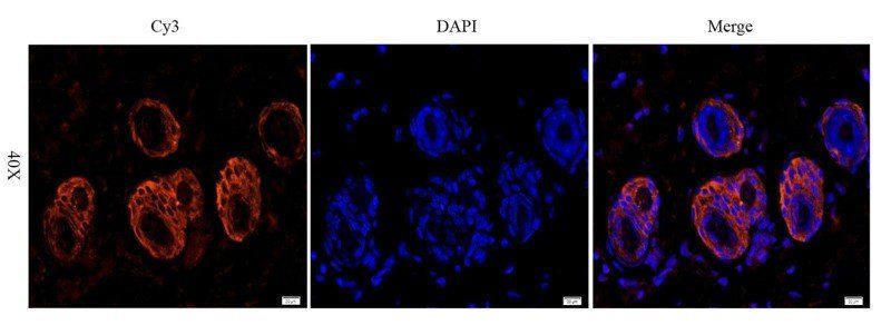
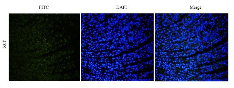
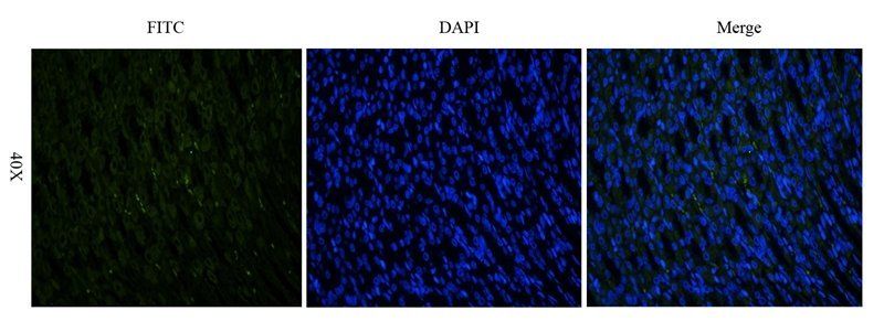
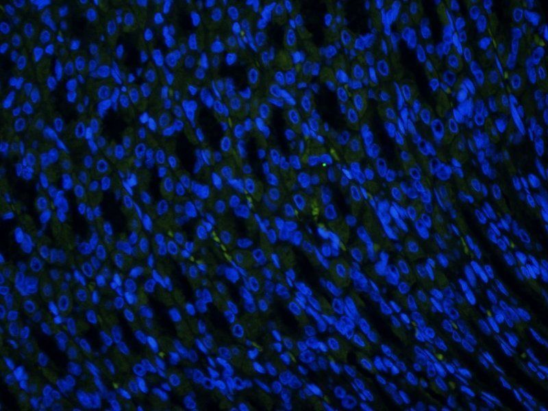
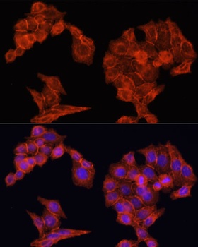
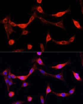
Immunofluorescent analysis of EGFR staining in HeLa cells. Formalin-fixed cells were permeabilized with 0.1% Triton X-100 in TBS for 5-10 minutes and blocked with 3% BSA-PBS for 30 minutes at room temperature. Cells were probed with the primary antibody in 3% BSA-PBS and incubated overnight at 4 °C in a hidified chamber. Cells were washed with PBST and incubated with a DyLight 594-conjugated secondary antibody (red) in PBS at room temperature in the dark. DAPI was used to stain the cell nuclei (blue).
- Item 1 of 18
EGFR isoform a variant antibody [orb308736]
ELISA, ICC, IF, IHC-P, WB
Human, Mouse, Porcine, Rat
Rabbit
Polyclonal
Unconjugated
100 μg, 200 μg - Item 1 of 8
- Item 1 of 9
- Item 1 of 7
- Item 1 of 8

Submit a review
Filter by Rating

- 5 stars

- 4 stars
- 3 stars
- 2 stars
- 1 stars
