You have no items in your shopping cart.
Cart summary
Item 1 of 7
Item 1 of 7
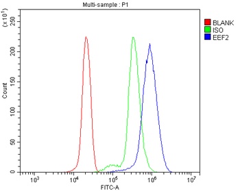
EEF2 Antibody
Catalog Number: orb692171
| Catalog Number | orb692171 |
|---|---|
| Category | Antibodies |
| Description | EEF2 Antibody |
| Species/Host | Rabbit |
| Clonality | Polyclonal |
| Tested applications | ELISA, IHC, WB |
| Reactivity | Human, Monkey, Mouse, Rat, Zebrafish |
| Isotype | Rabbit IgG |
| Immunogen | E.coli-derived human EEF2 recombinant protein (Position: M1-L858). |
| Concentration | Adding 0.2 ml of distilled water will yield a concentration of 500 μg/ml. |
| Dilution range | Western blot, 0.1-0.25μg/ml, Human, Monkey, Mouse, Rat, Zebrafish Immunohistochemistry (Paraffin-embedded Section), 2-5μg/ml, Human, Mouse, Rat Direct ELISA, 0.1-0.5μg/ml, Human |
| Form/Appearance | Lyophilized |
| Conjugation | Unconjugated |
| MW | 95 kDa |
| UniProt ID | P13639 |
| Storage | Store at -20˚C for one year from date of receipt. After reconstitution, at 4˚C for one month. It can also be aliquotted and stored frozen at -20˚C for six months. Avoid repeated freeze-thaw cycles. |
| Note | For research use only |
| Application notes | Tested Species: In-house tested species with positive results. Other applications have not been tested. Optimal dilutions should be determined by end users. Add 0.2ml of distilled water will yield a concentration of 500μg/ml. |
| Expiration Date | 12 months from date of receipt. |
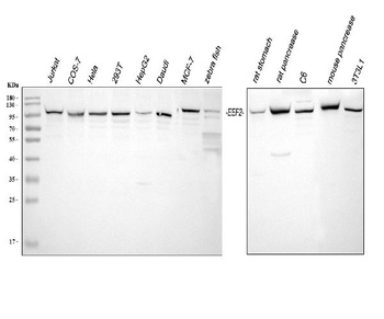
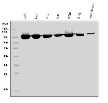
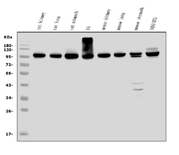
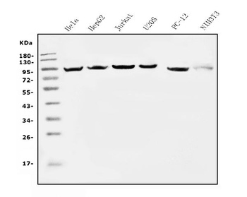
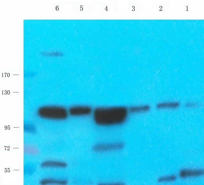
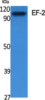
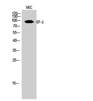
Western blot analysis of EEF2 using anti-EEF2 antibody (orb692171). Electrophoresis was performed on a 5-20% SDS-PAGE gel at 70V (Stacking gel)/90V (Resolving gel) for 2-3 hours. The sample well of each lane was loaded with 30 ug of sample under reducing conditions. Lane 1: human Jurkat whole cell lysates, Lane 2: monkey COS-7 whole cell lysates, Lane 3: human HeLa whole cell lysates, Lane 4: human 293T whole cell lysates, Lane 5: human HepG2 whole cell lysates, Lane 6: human Daudi whole cell lysates, Lane 7: human MCF-7 whole cell lysates, Lane 8: zebrafish tissue lysates, Lane 9: rat stomach tissue lysates, Lane 10: rat pancrease tissue lysates, Lane 11: rat C6 whole cell lysates, Lane 12: mouse pancrease tissue lysates, Lane 13: mouse 3T3L1 whole cell lysates. After electrophoresis, proteins were transferred to a nitrocellulose membrane at 150 mA for 50-90 minutes. Blocked the membrane with 5% non-fat milk/TBS for 1.5 hour at RT. The membrane was incubated with rabbit anti-EEF2 antigen affinity purified polyclonal antibody (Catalog # orb692171) at 0.25 μg/mL overnight at 4°C, then washed with TBS-0.1%Tween 3 times with 5 minutes each and probed with a goat anti-rabbit IgG-HRP secondary antibody at a dilution of 1:5000 for 1.5 hour at RT. The signal is developed using an Enhanced Chemiluminescent detection (ECL) kit (Catalog # orb90503) with Tanon 5200 system. A specific band was detected for EEF2 at approximately 95 kDa. The expected band size for EEF2 is at 95 kDa.
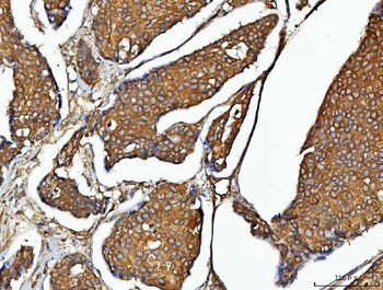
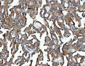
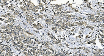
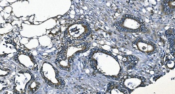
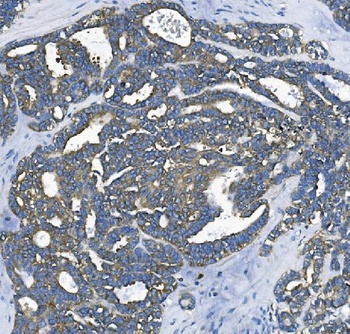
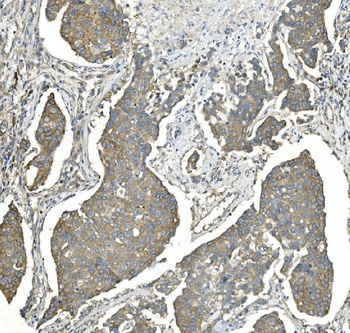
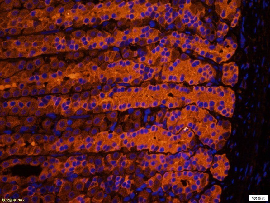
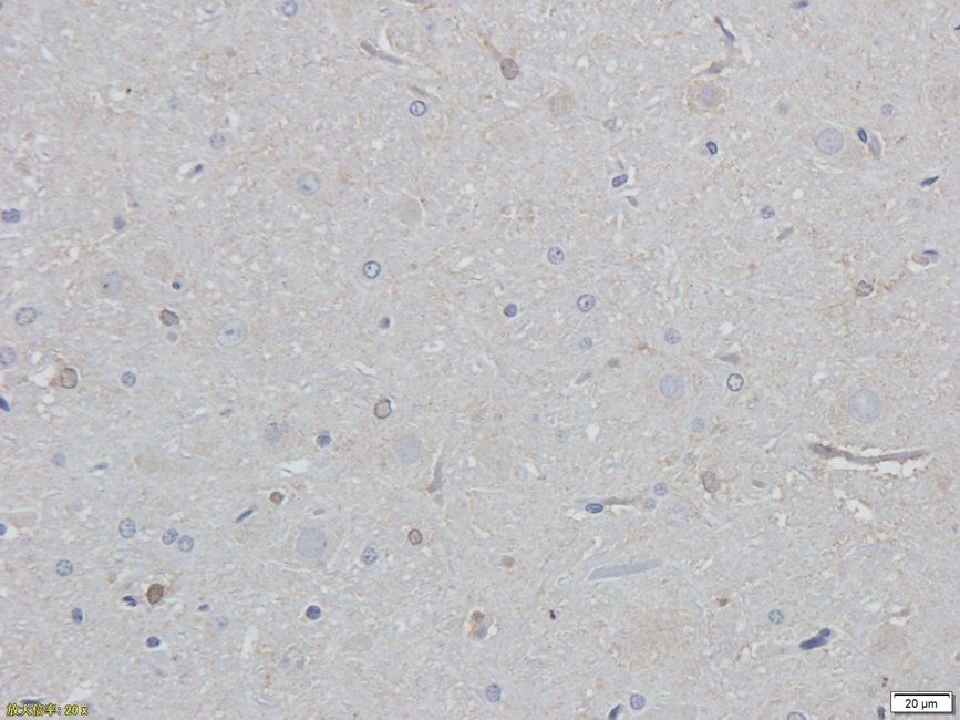
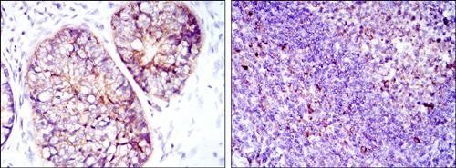
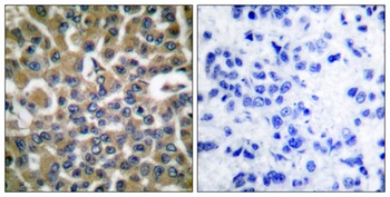
IHC analysis of EEF2 using anti-EEF2 antibody (orb692171). EEF2 was detected in a paraffin-embedded section of human breast cancer tissue. Heat mediated antigen retrieval was performed in EDTA buffer (pH 8.0, epitope retrieval solution). The tissue section was blocked with 10% goat serum. The tissue section was then incubated with 2 μg/ml rabbit anti-EEF2 Antibody (orb692171) overnight at 4°C. Peroxidase Conjugated Goat Anti-rabbit IgG was used as secondary antibody and incubated for 30 minutes at 37°C. The tissue section was developed using HRP Conjugated Rabbit IgG Super Vision Assay Kit with DAB as the chromogen.
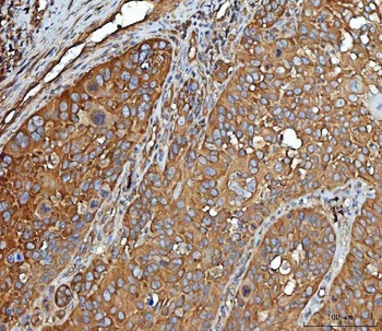
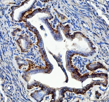
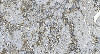
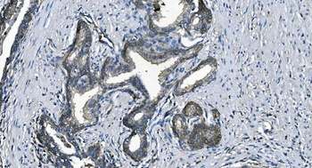
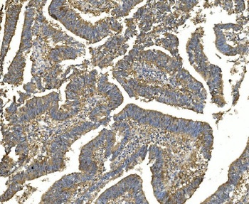
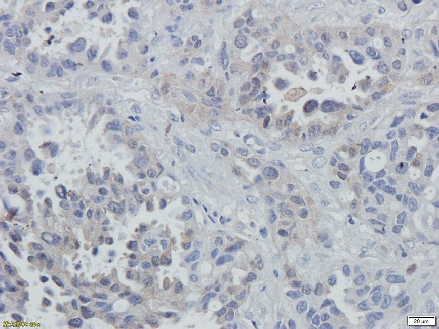
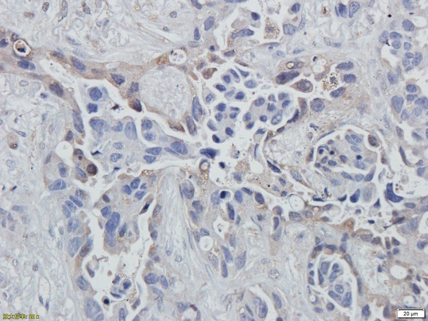
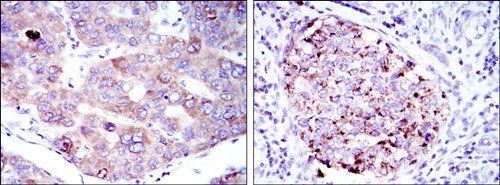
IHC analysis of EEF2 using anti-EEF2 antibody (orb692171). EEF2 was detected in a paraffin-embedded section of human laryngeal squamous cell carcinoma tissue. Heat mediated antigen retrieval was performed in EDTA buffer (pH 8.0, epitope retrieval solution). The tissue section was blocked with 10% goat serum. The tissue section was then incubated with 2 μg/ml rabbit anti-EEF2 Antibody (orb692171) overnight at 4°C. Peroxidase Conjugated Goat Anti-rabbit IgG was used as secondary antibody and incubated for 30 minutes at 37°C. The tissue section was developed using HRP Conjugated Rabbit IgG Super Vision Assay Kit with DAB as the chromogen.
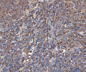
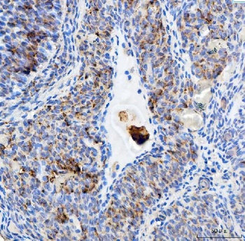
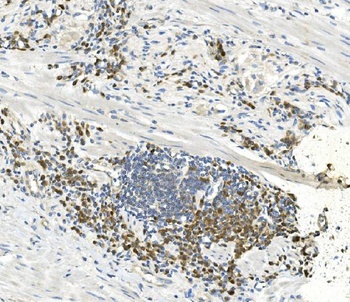
IHC analysis of EEF2 using anti-EEF2 antibody (orb692171). EEF2 was detected in a paraffin-embedded section of human lymphoma tissue. Heat mediated antigen retrieval was performed in EDTA buffer (pH 8.0, epitope retrieval solution). The tissue section was blocked with 10% goat serum. The tissue section was then incubated with 2 μg/ml rabbit anti-EEF2 Antibody (orb692171) overnight at 4°C. Peroxidase Conjugated Goat Anti-rabbit IgG was used as secondary antibody and incubated for 30 minutes at 37°C. The tissue section was developed using HRP Conjugated Rabbit IgG Super Vision Assay Kit with DAB as the chromogen.
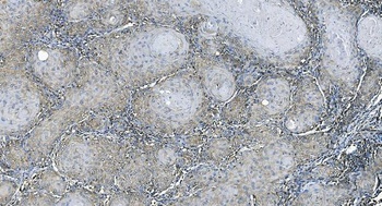
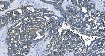
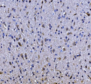
IHC analysis of EEF2 using anti-EEF2 antibody (orb692171). EEF2 was detected in a paraffin-embedded section of human renal cancer tissue. Heat mediated antigen retrieval was performed in EDTA buffer (pH 8.0, epitope retrieval solution). The tissue section was blocked with 10% goat serum. The tissue section was then incubated with 2 μg/ml rabbit anti-EEF2 Antibody (orb692171) overnight at 4°C. Peroxidase Conjugated Goat Anti-rabbit IgG was used as secondary antibody and incubated for 30 minutes at 37°C. The tissue section was developed using HRP Conjugated Rabbit IgG Super Vision Assay Kit with DAB as the chromogen.
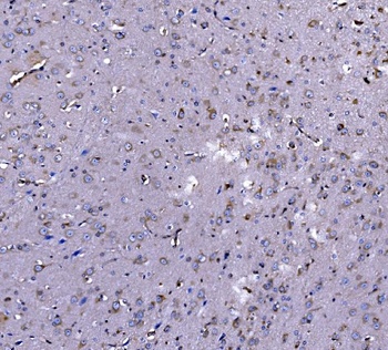
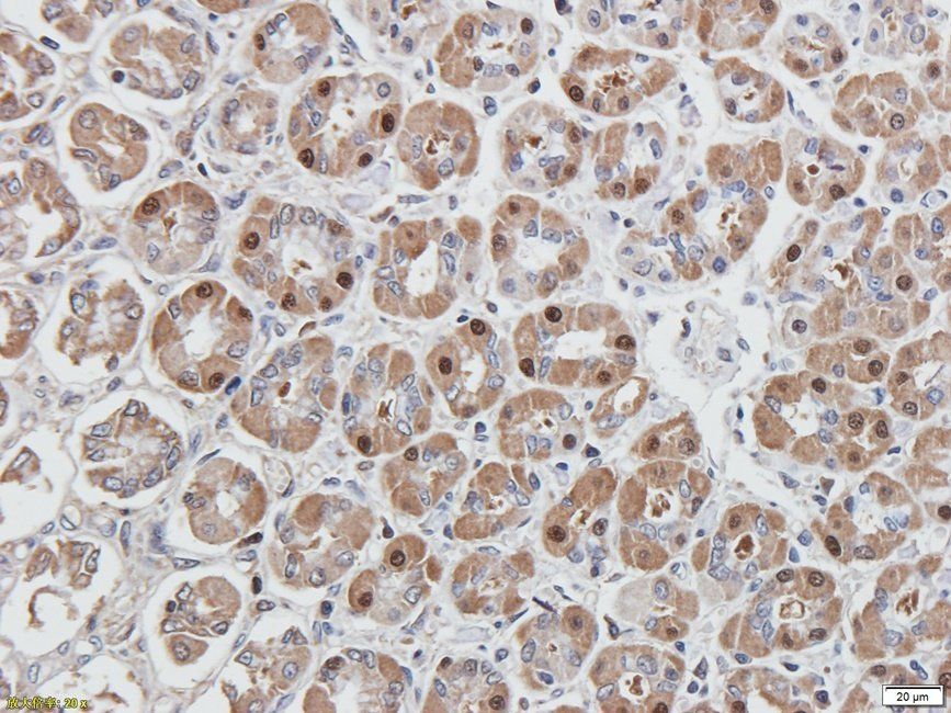
IHC analysis of EEF2 using anti-EEF2 antibody (orb692171). EEF2 was detected in a paraffin-embedded section of mouse ovary tissue. Heat mediated antigen retrieval was performed in EDTA buffer (pH 8.0, epitope retrieval solution). The tissue section was blocked with 10% goat serum. The tissue section was then incubated with 2 μg/ml rabbit anti-EEF2 Antibody (orb692171) overnight at 4°C. Peroxidase Conjugated Goat Anti-rabbit IgG was used as secondary antibody and incubated for 30 minutes at 37°C. The tissue section was developed using HRP Conjugated Rabbit IgG Super Vision Assay Kit with DAB as the chromogen.
IHC analysis of EEF2 using anti-EEF2 antibody (orb692171). EEF2 was detected in a paraffin-embedded section of rat ovary tissue. Heat mediated antigen retrieval was performed in EDTA buffer (pH 8.0, epitope retrieval solution). The tissue section was blocked with 10% goat serum. The tissue section was then incubated with 2 μg/ml rabbit anti-EEF2 Antibody (orb692171) overnight at 4°C. Peroxidase Conjugated Goat Anti-rabbit IgG was used as secondary antibody and incubated for 30 minutes at 37°C. The tissue section was developed using HRP Conjugated Rabbit IgG Super Vision Assay Kit with DAB as the chromogen.
- Item 1 of 13
EEF2/Elongation factor 2 Antibody [orb738382]
FC, ICC, IF, IHC, WB
Human, Monkey, Mouse, Rat
Rabbit
Polyclonal
Unconjugated
10 μg, 100 μg - Item 1 of 9
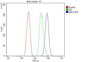
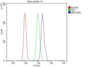
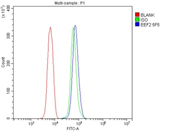
EEF2 Antibody (monoclonal, 5F5) [orb692233]
FC, ICC, IF, IHC, WB
Human, Mouse, Rat
Mouse
Monoclonal
Unconjugated
10 μg, 100 μg - Item 1 of 6
- Item 1 of 5
- Item 1 of 4
EF-2 antibody [orb765105]
ELISA, IF, IHC-P, WB
Human, Mouse, Rat
Rabbit
Polyclonal
Unconjugated
50ul, 100ul

Submit a review
Filter by Rating

- 5 stars

- 4 stars
- 3 stars
- 2 stars
- 1 stars