You have no items in your shopping cart.
Cart summary
Item 1 of 3
Item 1 of 3
E2F4 Antibody
Catalog Number: orb1825280

| Catalog Number | orb1825280 |
|---|---|
| Category | Antibodies |
| Description | The human retinoblastoma gene product appears to play an important role in the negative regulation of cell proliferation. Functional inactivation of Rb can be mediated either through mutation or as a consequence of interaction with DNA tumor virus-encoded proteins. Of all the Rb associations described to date, the identification of a complex between Rb and the transcription factor E2F most directly implicates Rb in regulation of cell proliferation. E2F was originally identified through its role in transcriptional activation of the adenovirus E2 promoter. Sequences homologous to the E2F binding site have been found upstream of a number of genes that encode proteins with putative functions in the G1 and S phases of the cell cycle. E2F-1 is a member of a broader family of transcription regulators including E2F-2, E2F-3, E2F-4, E2F-5, E2F-6 and E2F-7 each of which forms heterodimers with a second protein, DP-1, forming an active E2F transcriptional regulatory complex. |
| Species/Host | Mouse |
| Clonality | Monoclonal |
| Clone Number | E2F4/4224 |
| Tested applications | IHC-P |
| Reactivity | Human |
| Isotype | Mouse IgG1, kappa |
| Immunogen | A recombinant partial protein sequence (within amino acids 1-100) from the human protein was used as the immunogen for the E2F4 antibody. |
| Antibody Type | Primary Antibody |
| Dilution range | Immunohistochemistry (FFPE): 1-2ug/ml for 30 minutes at RT |
| Conjugation | Unconjugated |
| Formula | 1 mg/ml in 1X PBS; BSA free, sodium azide free |
| Hazard Information | This E2F4 antibody is available for research use only. |
| UniProt ID | Q16254 |
| Storage | Aliquot the E2F4 antibody and store frozen at -20°C or colder. Avoid repeated freeze-thaw cycles. |
| Note | For research use only |
| Expiration Date | 12 months from date of receipt. |

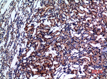
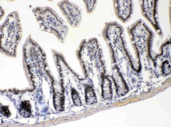
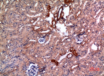
IHC staining of FFPE human lymph node tissue with E2F4 antibody (clone E2F4/4224). Inset: PBS used in place of primary Ab (secondary Ab negative control). HIER: boil tissue sections in pH9 10 mM Tris with 1 mM EDTA for 20 min and allow to cool before testing.
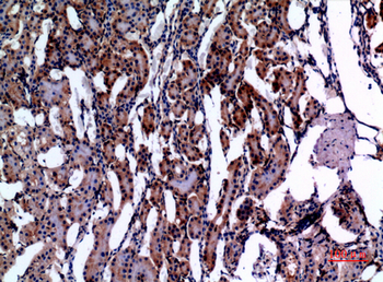
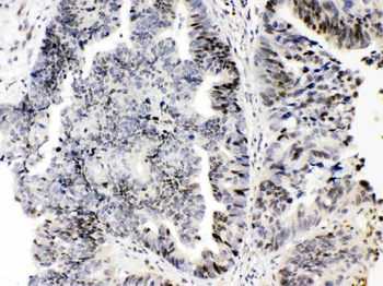
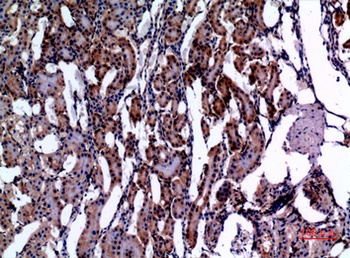
IHC staining of FFPE human breast carinoma tissue with E2F4 antibody (clone E2F4/4224). HIER: boil tissue sections in pH9 10 mM Tris with 1 mM EDTA for 20 min and allow to cool before testing.
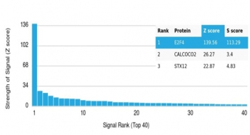
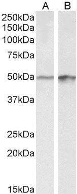
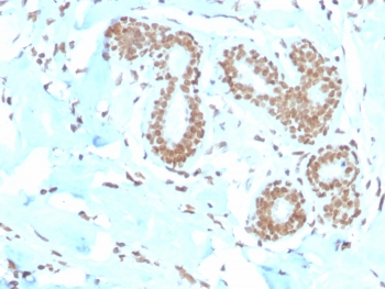
Analysis of a HuProt (TM) microarray containing more than 19000 full-length human proteins using E2F4 antibody (clone E2F4/4224). Z- and S- Score: The Z-score represents the strength of a signal that a monoclonal antibody (in combination with a fluorescently-tagged anti-IgG secondary antibody) produces when binding to a particular protein on the HuProt (TM) array. Z-scores are described in units of standard deviations (SD's) above the mean value of all signals generated on that array. If targets on HuProt (TM) are arranged in descending order of the Z-score, the S-score is the difference (also in units of SD's) between the Z-score. S-score therefore represents the relative target specificity of a mAb to its intended target. A mAb is considered to specific to its intended target, if the mAb has an S-score of at least 2.5. For example, if a mAb binds to protein X with a Z-score of 43 and to protein Y with a Z-score of 14, then the S-score for the binding of that mAb to protein X is equal to 29.
- Item 1 of 5
E2F-4 Polyclonal Antibody [orb1411448]
IHC-P, WB
Human, Mouse, Rat
Rabbit
Polyclonal
Unconjugated
100 μl - Item 1 of 2
Goat anti-Transcription factor E2F4 Antibody [orb146475]
ELISA, WB
Bovine, Canine, Human, Mouse, Porcine
Goat
Polyclonal
Unconjugated
100 μg - Item 1 of 3
E2F4 monoclonal antibody (M01), clone 5B7 [orb2293406]
ELISA, WB
Human
Mouse
Monoclonal
Unconjugated
100 μg - Item 1 of 4
- Item 1 of 4