You have no items in your shopping cart.
Cart summary
Item 1 of 4
Item 1 of 4
DBC1 Antibody / Deleted in breast cancer 1 / CCAR2
Catalog Number: orb1822897

| Catalog Number | orb1822897 |
|---|---|
| Category | Antibodies |
| Description | DBC-1 (deleted in breast cancer gene 1 protein), also known as p30 DBC protein and Cell cycle and apoptosis regulator protein 2 (CCAR2), is one of the genes located within the region of chromosome 8p21.3 that is homozygously deleted in some breast cancers. DBC-1 contains a nuclear localization signal, an N-terminal leucine zipper, an EF hand and a C-terminal coiled-coil region. DBC-1 is closely related to DIS but lacks the SAP domain. During death signaling mediated by TNF-Alpha, endogenous DBC-1 undergoes caspase-dependent processing to generate DBC-1 p120 and p66, both of which include the C-terminus of the protein. Both DBC-1 p120 and p66 relocate to the cytoplasm. Overexpression of the DBC-1 p120 form results in mitochondrial clustering and matrix condensation and increases the sensitivity of cells to TNF-Alpha-mediated apoptosis. In addition, DBC-1 directly interacts with unliganded ER-Alpha, stabilizing its expression and therefore collaborating to suppress apoptosis and promote hormone-independent cell growth. |
| Clonality | Monoclonal |
| Species/Host | Mouse |
| Isotype | Mouse IgG2a |
| Conjugation | Unconjugated |
| Reactivity | Human |
| Immunogen | Recombinant full-length human CCAR2 / KIAA1967 protein was used as the immunogen for the DBC1 antibody. |
| UniProt ID | Q8N163 |
| Tested applications | FACS, IHC-P |
| Dilution range | Flow cytometry: 1-2ug/million cells,Immunohistochemistry (FFPE): 1-2ug/ml for 30 min at RT |
| Antibody Type | Primary Antibody |
| Clone Number | PCRP-KIAA1967-1D10 |
| Formula | 0.2 mg/ml in 1X PBS with 0.1 mg/ml BSA (US sourced), 0.05% sodium azide |
| Storage | Aliquot the DBC1 antibody and store frozen at -20°C or colder. Avoid repeated freeze-thaw cycles. |
| Hazard Information | This DBC1 antibody is available for research use only. |
| Note | For research use only |
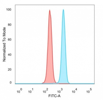
Flow cytometry testing of PFA-fixed human HeLa cells with DBC1 antibody (clone PCRP-KIAA1967-1D10) followed by goat anti-mouse IgG-CF488 (blue); Red = unstained cells.
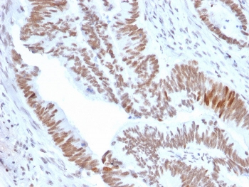
IHC staining of FFPE human colon tumor tissue with DBC1 antibody (clone PCRP-KIAA1967-1D10). HIER: boil tissue sections in pH9 10 mM Tris with 1 mM EDTA for 20 min and allow to cool before testing.

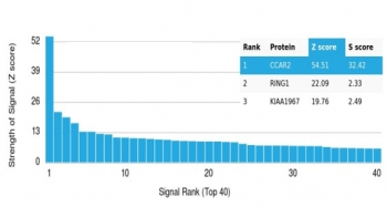
Analysis of a HuProt (TM) microarray containing more than 19000 full-length human proteins using DBC1 antibody (clone PCRP-KIAA1967-1D10). Z- and S- Score: The Z-score represents the strength of a signal that a monoclonal antibody (in combination with a fluorescently-tagged anti-IgG secondary antibody) produces when binding to a particular protein on the HuProt (TM) array. Z-scores are described in units of standard deviations (SD's) above the mean value of all signals generated on that array. If targets on HuProt (TM) are arranged in descending order of the Z-score, the S-score is the difference (also in units of SD's) between the Z-score. S-score therefore represents the relative target specificity of a mAb to its intended target. A mAb is considered to specific to its intended target, if the mAb has an S-score of at least 2.5. For example, if a mAb binds to protein X with a Z-score of 43 and to protein Y with a Z-score of 14, then the S-score for the binding of that mAb to protein X is equal to 29.

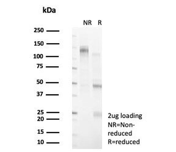
SDS-PAGE analysis of purified, BSA-free DBC1 antibody (clone PCRP-KIAA1967-1D10) as confirmation of integrity and purity.
- Item 1 of 4
DBC1 Antibody / Deleted in breast cancer 1 / CCAR2 [orb1822895]
FACS, IHC-P
Human
Mouse
Monoclonal
Unconjugated
100 μg - Item 1 of 4
DBC1 Antibody / Deleted in breast cancer 1 / CCAR2 [orb1822896]
FACS, IHC-P
Human
Mouse
Monoclonal
Unconjugated
20 μg