You have no items in your shopping cart.
Cart summary
Item 1 of 5
Item 1 of 5
DARS Antibody (N-term)
Catalog Number: orb1938464

| Catalog Number | orb1938464 |
|---|---|
| Category | Antibodies |
| Description | Purified Rabbit Polyclonal Antibody (Pab) |
| Target | This DARS antibody is generated from rabbits immunized with a KLH conjugated synthetic peptide between 154-183 amino acids from the N-terminal region of human DARS. |
| Clonality | Polyclonal |
| Species/Host | Rabbit |
| Isotype | Rabbit IgG |
| Conjugation | Unconjugated |
| Reactivity | Human |
| Form/Appearance | Purified polyclonal antibody supplied in PBS with 0.09% (W/V) sodium azide. This antibody is prepared by Saturated Ammonium Sulfate (SAS) precipitation followed by dialysis against PBS. |
| UniProt ID | P14868 |
| MW | 57136 Da |
| Tested applications | FC, IF, IHC-P, WB |
| Dilution range | IF: 1:10~50, WB: 1:1000, WB: 1:1000, IHC-P: 1:50~100, FC: 1:10~50 |
| Antibody Type | Primary Antibody |
| Clone Number | RB14801 |
| Storage | Maintain refrigerated at 2-8°C for up to 2 weeks. For long term storage store at -20°C in small aliquots to prevent freeze-thaw cycles |
| Alternative names | Aspartate--tRNA ligase, cytoplasmic, Aspartyl-tRNA Read more... |
| Note | For research use only |
| NCBI | NP_001340.2 |
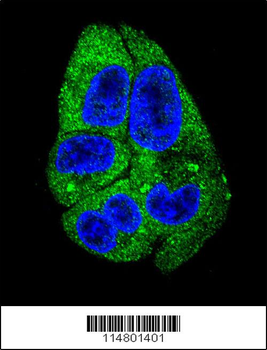
Confocal immunofluorescent analysis of DARS Antibody (N-term) with HepG2 cell followed by Alexa Fluor 488-conjugated goat anti-rabbit lgG (green). DAPI was used to stain the cell nuclear (blue).
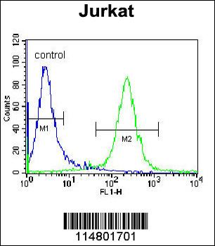
DARS Antibody (N-term) flow cytometric analysis of Jurkat cells (right histogram) compared to a negative control cell (left histogram). FITC-conjugated donkey-anti-rabbit secondary antibodies were used for the analysis.
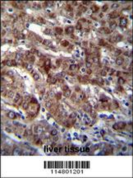
DARS Antibody (N-term) immunohistochemistry analysis in formalin fixed and paraffin embedded human liver tissue followed by peroxidase conjugation of the secondary antibody and DAB staining. This data demonstrates the use of DARS Antibody (N-term) for immunohistochemistry. Clinical relevance has not been evaluated.
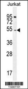
DARS Antibody (N-term) western blot analysis in Jurkat cell line lysates (35 ug/lane). This demonstrates the DARS antibody detected the DARS protein (arrow).
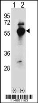
Western blot analysis of DARS (arrow) using rabbit polyclonal DARS Antibody (N-term). 293 cell lysates (2 ug/lane) either nontransfected (Lane 1) or transiently transfected (Lane 2) with the DARS gene.
