You have no items in your shopping cart.
Cart summary
Item 1 of 4
Item 1 of 4
CTBP1 Antibody
Catalog Number: orb1267852
| Catalog Number | orb1267852 |
|---|---|
| Category | Antibodies |
| Description | CTBP1 Antibody |
| Species/Host | Rabbit |
| Clonality | Polyclonal |
| Tested applications | IF, WB |
| Reactivity | Human, Mouse, Rat |
| Isotype | Rabbit Ig |
| Immunogen | This CTBP1 antibody is generated from rabbits immunized with a KLH conjugated synthetic peptide between 413-440 amino acids from the C-terminal region of human CTBP1. |
| Concentration | batch dependent |
| Dilution range | For WB starting dilution is: 1:1000For IF starting dilution is: 1:10~50 |
| Form/Appearance | Liquid |
| Conjugation | Unconjugated |
| MW | 48 kDa |
| Target | CTBP1 |
| UniProt ID | Q13363 |
| NCBI | Q13363 |
| Storage | Store at 4°C for three months and -20°C, stable for up to one year. As with all antibodies care should be taken to avoid repeated freeze thaw cycles. Antibodies should not be exposed to prolonged high temperatures. |
| Buffer/Preservatives | Supplied in PBS with 0.09% (W/V) sodium azide. |
| Alternative names | C-terminal-binding protein 1, CtBP1, 111-, CTBP1, Read more... |
| Note | For research use only |
| Application notes | For WB starting dilution is: 1:1000For IF starting dilution is: 1:10~50 |
| Expiration Date | 12 months from date of receipt. |
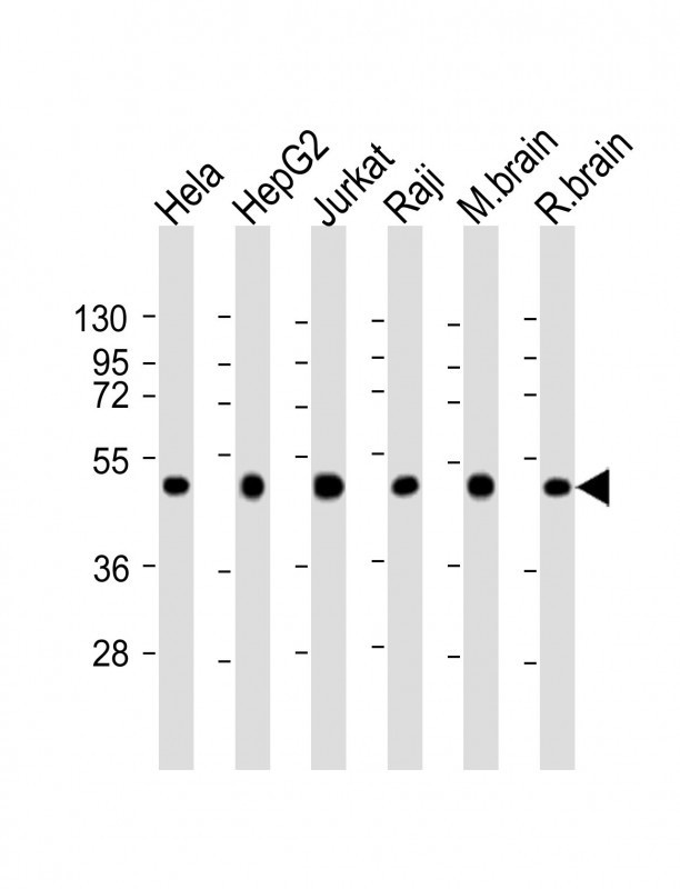
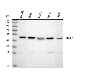
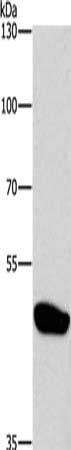
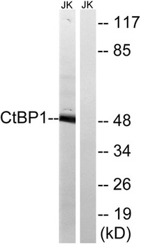
Western Blot at 1:2000 dilution Lane 1: Hela whole cell lysate Lane 2: HepG2 whole cell lysate Lane 3: Jurkat whole cell lysate Lane 4: Raji whole cell lysate Lane 5: mouse brain lysate Lane 6: rat brain lysate Lysates/proteins at 20 ug per lane.
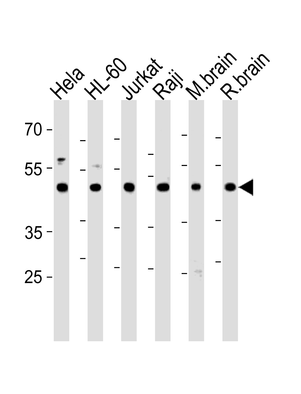
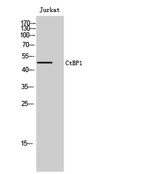
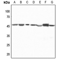
Western blot analysis in Hela, HL-60, Jurkat, Raji cell line and mouse brain, rat brain tissue lysates (35 ug/lane).
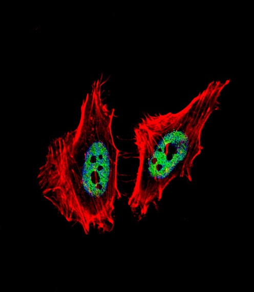
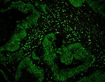
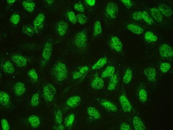
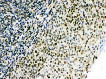
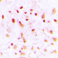
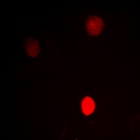
Fluorescent confocal image of Hela cell stained with CTBP1 Antibody. Hela cells were fixed with 4% PFA (20 min), permeabilized with Triton X-100 (0.1%, 10 min), then incubated with CTBP1 primary antibody (1:25). For secondary antibody, Alexa Fluor 488 conjugated donkey anti-rabbit antibody (green) was used (1:400). Cytoplasmic actin was counterstained with Alexa Fluor 555 (red) conjugated Phalloidin (7 units/ml). Nuclei were counterstained with DAPI (blue) (10 ug/ml, 10 min).CTBP1 immunoreactivity is localized to nucleus significantly.
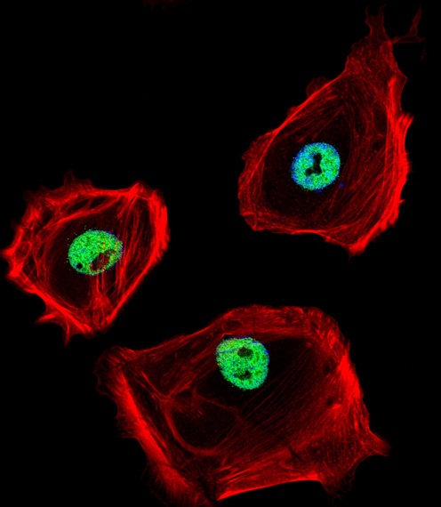
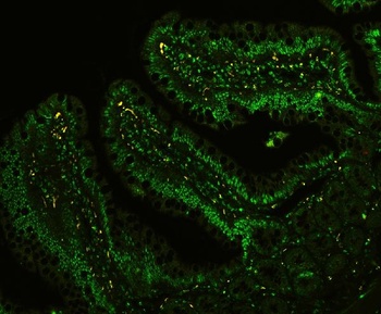
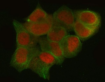
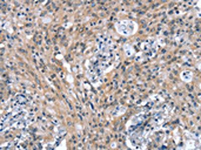
Fluorescent confocal image of SK-BR-3 cell stained with CTBP1 Antibody. SK-BR-3 cells were fixed with 4% PFA (20 min), permeabilized with Triton X-100 (0.1%, 10 min), then incubated with CTBP1 primary antibody (1:25). For secondary antibody, Alexa Fluor 488 conjugated donkey anti-rabbit antibody (green) was used (1:400). Cytoplasmic actin was counterstained with Alexa Fluor 555 (red) conjugated Phalloidin (7 units/ml). Nuclei were counterstained with DAPI (blue) (10 ug/ml, 10 min).CTBP1 immunoreactivity is localized to nucleus significantly.
- Item 1 of 9
CtBP1 Antibody [orb259602]
FC, ICC, IF, IHC, WB
Hamster
Human, Mouse, Rat
Rabbit
Polyclonal
Unconjugated
10 μg, 100 μg - Item 1 of 3
CTBP1 antibody [orb524034]
ELISA, IHC, WB
Human, Mouse, Rat
Rabbit
Polyclonal
Unconjugated
50 μl, 100 μl - Item 1 of 3
CTBP1 antibody [orb524033]
ELISA, IHC, WB
Human, Mouse, Rat
Rabbit
Polyclonal
Unconjugated
50 μl, 100 μl - Item 1 of 4
CtBP1 antibody [orb767758]
ELISA, IHC-P, WB
Human, Mouse, Rat
Rabbit
Polyclonal
Unconjugated
50ul, 100ul - Item 1 of 3
CTBP1 antibody [orb256481]
IF, IH, WB
Human, Mouse, Porcine, Rat
Rabbit
Polyclonal
Unconjugated
200 μl, 100 μl, 30 μl


Submit a review
Filter by Rating

- 5 stars

- 4 stars
- 3 stars
- 2 stars
- 1 stars