You have no items in your shopping cart.
Cart summary
Item 1 of 3
Item 1 of 3
COXI Antibody (Center)
Catalog Number: orb1927786

| Catalog Number | orb1927786 |
|---|---|
| Category | Antibodies |
| Description | Affinity Purified Rabbit Polyclonal Antibody (Pab) |
| Target | This COXI antibody is generated from rabbits immunized with a KLH conjugated synthetic peptide between 195-224 amino acids of human COXI. |
| Clonality | Polyclonal |
| Species/Host | Rabbit |
| Isotype | Rabbit IgG |
| Conjugation | Unconjugated |
| Reactivity | Human |
| Predicted Reactivity | Equine, Mouse, Other, Porcine, Rabbit, Rat, Sheep, Zebrafish |
| Form/Appearance | Purified polyclonal antibody supplied in PBS with 0.09% (W/V) sodium azide. This antibody is purified through a protein A column, followed by peptide affinity purification. |
| UniProt ID | P00395 |
| MW | 57041 Da |
| Tested applications | IF, WB |
| Dilution range | IF: 1:25, IF: 1:25, WB: 1:2000 |
| Antibody Type | Primary Antibody |
| Clone Number | RB24524 |
| Storage | Maintain refrigerated at 2-8°C for up to 2 weeks. For long term storage store at -20°C in small aliquots to prevent freeze-thaw cycles |
| Alternative names | Cytochrome c oxidase subunit 1, Cytochrome c oxida Read more... |
| Note | For research use only |
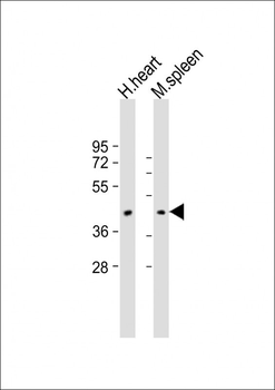
All lanes: Anti-COXI Antibody (Center) at 1:2000 dilution. Lane 1: human heart lysate. Lane 2: mouse spleen lysate. Lysates/proteins at 20 µg per lane. Secondary Goat Anti-Rabbit IgG, (H+L), Peroxidase conjugated at 1/10000 dilution. Predicted band size: 57 kDa. Blocking/Dilution buffer: 5% NFDM/TBST.
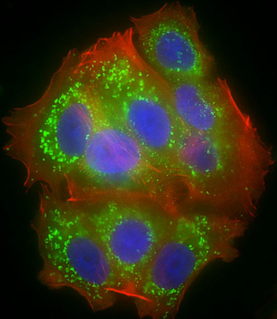
Immunofluorescent analysis of 4% paraformaldehyde-fixed, 0.1% Triton X-100 permeabilized MCF-7 (human breast cancer cell line) cells labeling COXI at 1/25 dilution, followed by Dylight 488-conjugated goat anti-rabbit IgG secondary antibody at 1/200 dilution (green). Immunofluorescence image showing cytoplasm and mitochondrion staining on MCF-7 cell line. Cytoplasmic actin is detected with Dylight 554 Phalloidin at 1/100 dilution (red). The nuclear counter stain is DAPI (blue).
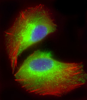
Immunofluorescent analysis of 4% paraformaldehyde-fixed, 0.1% Triton X-100 permeabilized U-2 OS (human osteosarcoma cell line) cells labeling COXI at 1/25 dilution, followed by Dylight 488-conjugated goat anti-rabbit IgG (1583138) secondary antibody at 1/200 dilution (green). Immunofluorescence image showing endoplasmic reticulum staining on U-2 OS cell line. Cytoplasmic actin is detected with Dylight 554 Phalloidin at 1/100 dilution (red).The nuclear counter stain is DAPI (blue).
